Chapter 16 Mediastinal Masses
DIAGNOSTIC IMAGING WORKUP OF MEDIASTINAL MASSES
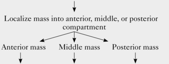
Radiologic examination of a mediastinal mass usually can narrow the differential diagnosis to two or three likely candidates. In some cases, imaging features enable the radiologist to make a specific diagnosis. The radiologic workup depends on the location of the mass (Fig. 16-1 and Box 16-1).

Figure 16-1 Mediastinal compartments. A lateral chest radiograph demonstrates the boundaries of the anterior, middle, and posterior mediastinal compartments.
Box 16-1 Algorithm for Imaging Evaluation of Mediastinal Masses
Mediastinal mass detected on chest radiograph | ||
![]() | ||
Chest CT | Chest CT (I+)* | MRI or Barium swallow if esophageal origin is suspected |
Other studies:Gallium scan if Hodgkin’s lymphoma is suspectedRadioiodine scanif thyroid goiter issuspected | Other studies:MRI if there is acontraindicationto contrast and avascular abnormalityis suspected or ifvascular invasionis suspected |
For masses localized within the anterior compartment of the mediastinum, computed tomography (CT) is a good diagnostic choice. It provides information about the precise location of a mass and its relation to adjacent mediastinal structures. CT can also determine whether a mass is cystic or solid and whether it contains calcium or fat. In many cases, non–contrast-enhanced CT is sufficient, but in others, contrast-enhanced CT can provide important information concerning enhancement of the mass and its relation to adjacent vascular structures. Correlative nuclear medicine studies may help in suspected cases of Hodgkin’s lymphoma (i.e., gallium scan) and substernal goiter (i.e., radioiodine scan).
CT is the preferred imaging modality for further evaluation of a middle mediastinal mass. Contrast-enhanced CT is preferred for the evaluation of middle mediastinal masses, especially when you suspect a vascular abnormality. The development of multidetector-row spiral CT scanners has improved the ability to evaluate mediastinal vascular abnormalities. Compared with single-detector spiral CT scanners, multidetector-row CT scanners are associated with improved spatial resolution, faster scanning, improved vascular enhancement, and higher-quality multiplanar reformation images.
Magnetic resonance imaging (MRI) has historically been considered superior to contrast-enhanced CT in assessing relationships of masses and vascular structures and in determining vascular invasion. However, in the era of multidetector-row CT scanners, there is less of a distinction between these two modalities for assessing these parameters, especially when a CT study is specifically tailored to evaluate vascular structures. MRI should be the first cross-sectional imaging study for patients with a suspected vascular abnormality who have a contraindication to intravenous contrast. MRI also plays an important second-line role to CT by providing further tissue characterization in cases in which a mass is incompletely characterized by CT.
For masses localized within the posterior mediastinum, MRI is usually preferred because of its superior ability to assess the relationship of the mass to the adjacent spine. An exception occurs when a posterior mediastinal mass is suspected to be of esophageal origin. In this situation, a barium swallow should be obtained.
The algorithm presented in this chapter is a general guideline. The decision to obtain CT or MRI data depends on several factors, including the availability of MRI, patient factors (contraindication to intravenous contrast or MRI), and institutional practices. In some cases, CT and MRI provide complementary information, and both may be indicated.
ANTERIOR MEDIASTINAL MASSES
Thymic Abnormalities
The thymus is a bilobed structure that is normally located within the anterior mediastinum. The thymus reaches its maximum weight at puberty and subsequently undergoes fatty involution over a 5- to 15-year period. Many thymic abnormalities can manifest as an anterior mediastinal mass, but the most common are thymic hyperplasia and thymic epithelial tumors.
Thymic Epithelial Tumors
The most widely accepted classification scheme for thymic epithelial neoplasms is the World Health Organization (WHO) histologic classification that was published in 1999 and updated in 2004. It divides thymic epithelial neoplasms into three main groups: low-risk thymomas (types A, AB, and B1), high-risk thymomas (types B2 and B3), and thymic carcinomas, including neuroendocrine epithelial tumors (type C).
The most common thymic abnormality that manifests as an anterior mediastinal mass is thymoma (Box 16-2). Thymomas account for most anterior mediastinal masses in adults and typically occur as incidental findings in otherwise healthy individuals. However, thymomas may be associated with other abnormalities, including myasthenia gravis, red cell aplasia, hypogammaglobulinemia, and stiff-person syndrome. The association with myasthenia gravis is the most common of the three; about 15% of patients with myasthenia gravis have a thymoma, and about 50% of patients with thymomas have myasthenia gravis.
DEMOGRAPHICS
Age: usually 40 to 60 years old; unusual in patients younger than 30 years old
Gender: men and women equally affected
Associations: myasthenia gravis, hypogammaglobulinemia, red cell aplasia, and stiff-person syndrome
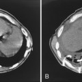
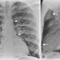
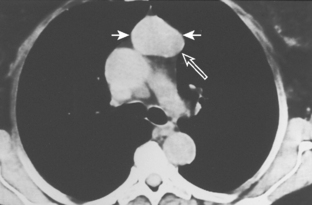
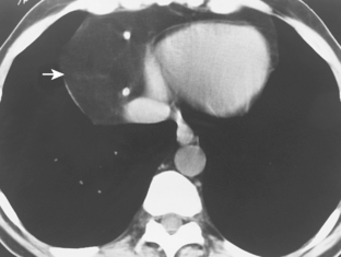
As with other midline anterior mediastinal masses, conventional radiographic findings are often limited to the lateral chest radiograph, which may demonstrate a well-defined mass in the normally clear retrosternal space. Small masses may not be detectable on plain radiographs, but CT can help to identify a thymoma in patients with myasthenia gravis. Characteristic CT imaging features include a well-defined, round, or oval mass, usually of homogeneous soft tissue density, that is located within the anterior mediastinum (Fig. 16-2). Although they are most commonly located anterior to the junction of the heart and great vessels, thymomas may occur at any level from the thoracic inlet to the diaphragm. In about 20% of cases, there is evidence of calcification, which is typically curvilinear.

Figure 16-2 Thymoma. Axial CT shows an oval, homogeneous soft tissue mass (short solid arrows) in the anterior mediastinum, with a thin rim of peripheral calcification posteriorly (open arrow). Calcification occurs in approximately 20% of thymomas.
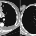
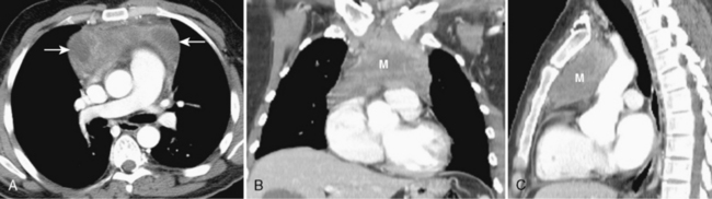
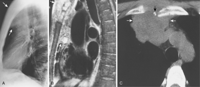
Most thymomas are benign lesions confined within a fibrous capsule, but about 30% of thymomas are more aggressive and demonstrate invasion through the fibrous capsule. The WHO classification scheme correlates with the likelihood of invasiveness, a factor that has an important influence on treatment and prognosis. Types A and AB are usually encapsulated, type B (especially B3) has a greater likelihood of invasiveness, and type C is almost always invasive. Although CT and MRI findings are often of limited value in differentiating histologic subtypes of thymic epithelial neoplasms, certain findings have predictive value. For example, a small, smooth, round, homogeneous anterior mediastinal mass usually corresponds to a type A thymoma, whereas irregular contours and heterogeneous attenuation favor a type C thymic carcinoma (Fig. 16-3). Calcification is most commonly observed in type B thymomas and type C thymic carcinoma.

Figure 16-3 Thymic carcinoma. Axial, contrast-enhanced CT (A) at the level of main pulmonary artery shows a large, heterogeneously enhancing, anterior mediastinal mass with foci of low attenuation (arrows), consistent with necrosis. Coronal (B) and sagittal (C) reformation images improve the assessment of the craniocaudal extent of the neoplastic mass (M) compared with axial images.
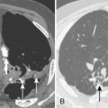
When interpreting CT scans or MRI studies of patients with suspected or proven thymic neoplasms, signs of capsular invasion or extracapsular extension should be carefully sought. They include irregular tumor margins; invasion of surrounding mediastinal fat, vascular structures, or chest wall; and irregular interface with the adjacent lung. Invasive thymomas typically spread locally (Fig. 16-4), and metastases outside of the thorax are rare. Pleural dissemination, also referred to as drop metastases, and pericardial involvement are common, whereas lung metastases are rare. The extent of involvement by thymic neoplasms is often best determined by viewing CT or MRI data in axial, sagittal, and coronal planes rather than relying solely on axial images (see Fig. 16-3).

Figure 16-4 Invasive thymoma. A, The posteroanterior chest radiograph shows a lobulated, anterior mediastinal mass (white arrows) and multiple pleural masses in the right hemithorax (open arrows). B, Axial CT image of the chest (filmed using soft tissue windows) shows an anterior mediastinal mass (black arrows) and multiple, unilateral pleural masses (curved arrows). Notice that one of the pleural masses is located within the minor fissure (open arrow). C, Axial CT image of the chest (filmed using lung windows) shows multiple pleural masses (curved arrows), including a pleural mass within the minor fissure (open arrow). The presence of an anterior mediastinal mass and unilateral pleural masses strongly suggests invasive thymoma.
Thymic Hyperplasia
Thymic hyperplasia (Box 16-3) is associated with a wide variety of systemic abnormalities, including hyperthyroidism. Rebound thymic hyperplasia may be seen in patients who have been treated with chemotherapy.
Thymic hyperplasia is usually identified on CT as enlargement of the thymus gland, which maintains its normal bilobed, arrowhead configuration (Fig. 16-5). In contrast, most other thymic abnormalities appear as a discrete mass rather than as uniform glandular enlargement. 18F-fluorodeoxyglucose positron emission tomography (FDG-PET) is of limited value in differentiating thymic hyperplasia from thymic neoplasms because both may demonstrate FDG avidity. MRI imaging using a chemical shift technique can reliably differentiate thymic hyperplasia from thymic neoplasms. On chemical shift imaging, thymic hyperplasia is associated with a characteristic decrease in signal intensity within the thymus gland.

Figure 16-5 Thymic hyperplasia caused by hyperthyroidism. Axial CT image of the chest at the level of the aortic arch (AA) shows enlargement of the thymus gland (arrows), which maintains its normal bilobed, arrowhead configuration.
Thymic lymphoid hyperplasia is a distinct entity that is characterized by an increased number of lymphoid follicles, but the gland is usually not enlarged. Thymic lymphoid hyperplasia is most commonly associated with myasthenia gravis. On imaging studies, the thymus gland usually appears normal, but it may uncommonly may appear as a focal mass or diffuse glandular enlargement.
Thymic Carcinoid
Thymic carcinoid tumor is rare and is thought to arise from thymic cells of neural crest origin. It is now included in the same histologic category as thymic carcinomas in the 2004 WHO classification of thymic epithelial neoplasms.
Patients with thymic carcinoid tumors often present with endocrine abnormalities, including Cushing’s syndrome, the syndrome of inappropriate antidiuretic hormone secretion (SIADH), hyperparathyroidism, and multiple endocrine neoplasia (MEN 1) syndrome.
No radiologic features differentiate thymic carcinoid from thymic carcinoma. The diagnosis is often suspected on the basis of endocrine abnormalities in a patient with an anterior mediastinal mass.
Thymic Cyst
Thymic cysts may be congenital or acquired. Acquired thymic cysts are most often associated with Hodgkin’s disease after radiation therapy.
Radiographically, a thymic cyst usually appears as a well-defined, cystic mass with an imperceptible wall. The CT attenuation values are typically consistent with fluid; however, the appearance may vary if hemorrhage or infection complicates the cyst. Curvilinear calcification of the cyst wall occurs in a few cases.
Thymic Lymphoma
Thymic involvement may occur in up to one third of patients with Hodgkin’s lymphoma. It is usually accompanied by involvement of mediastinal lymph nodes.
Thymic lymphoma usually manifests as a homogeneous, round mass with soft tissue density and without calcification. The presence of enlarged mediastinal lymph nodes may suggest the diagnosis.
Thymolipoma
Thymolipoma is a rare, benign thymic neoplasm composed primarily of fat, but it also contains strands of thymic tissue. Thymolipomas most often occur in younger patients and are usually identified on chest radiographs as incidental findings.
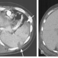
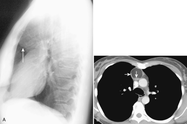
Because of their soft, pliable nature, thymolipomas commonly drape around the heart and other mediastinal structures. They are often quite large at the time of presentation, and they may mimic cardiac enlargement on chest radiographs. Identification of fat within the mass on CT or MRI suggests the diagnosis (Fig. 16-6).
Primary Mediastinal Lymphoma
Primary mediastinal lymphoma (Box 16-4) refers to malignant lymphoma that is exclusively or mostly limited to the mediastinum. The most common cell types to arise in the anterior mediastinum include the nodular sclerosing subtype of Hodgkin’s lymphoma, primary mediastinal large B-cell lymphoma, and lymphoblastic lymphoma.
Box 16-4 Primary Mediastinal Lymphoma
DEMOGRAPHICS
Age: mostly young adults, but Hodgkin’s lymphoma has a bimodal distribution, with initial peak in young adults and a second peak after age 50 years
Gender: female predominance, except for lymphoblastic lymphoma
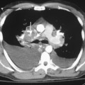
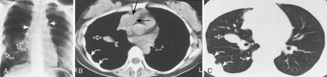
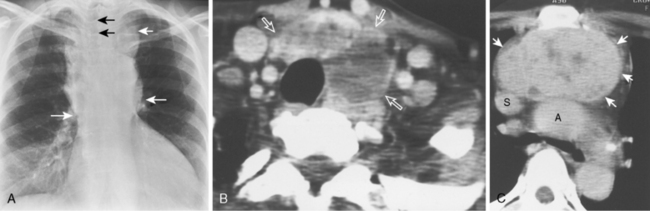
Approximately 40% of these patients present with enlargement of a single anterior mediastinal nodal group. Imaging features vary, ranging from a single, spherical soft tissue mass in the anterior mediastinum to a large, lobulated mass representing a conglomeration of lymph nodes. CT may show a mass with homogeneous soft tissue density, or it may appear heterogeneous, in which the low-attenuation areas represent necrosis. Whereas anterior mediastinal masses from lymphoma typically demonstrate well-defined margins, invasion of adjacent lung parenchyma may result in irregular margins. Invasion into the chest wall may also occur (Fig. 16-7).

Figure 16-7 Lymphoma. A, The lateral chest radiograph reveals increased opacity in the normally clear retrosternal space (lower arrow) and a presternal soft tissue mass (upper arrow). B, Sagittal MRI confirms the presence of a heterogeneous anterior mediastinal mass (long arrow) with invasion of the chest wall and extension into the presternal soft tissues (short arrow). C, Axial noncontrast CT scan shows destruction of the sternum (black arrow) by the anterior mediastinal mass (white arrows).
Lymphoma that manifests as a solitary, spherical mass may be indistinguishable from thymoma. Correlative nuclear medicine gallium imaging may be helpful because most Hodgkin’s lymphomas take up gallium avidly.
Associated lymphadenopathy in other compartments of the mediastinum and associated extrathoracic lymphadenopathy each suggest the diagnosis of lymphoma. An important discriminating feature is the absence of calcification in untreated lymphoma. Although the presence of calcification strongly suggests a diagnosis other than lymphoma, calcification frequently occurs in cases of treated lymphoma, but only rarely in untreated cases.
FDG-PET and FDG-PET/CT play important roles in staging lymphoma and in assessing the response to therapy. After treatment, patients often have residual mediastinal masses. PET is especially helpful in this setting because it can differentiate a residual, benign, fibrotic tissue mass from incompletely treated, viable tumor.
Germ Cell Neoplasms
Primary germ cell neoplasms (GCNs) arise from rests of primitive germ cells that were left within the mediastinum during their migration from the yolk sac to the urogenital ridge. The anterior mediastinum is the most common extragonadal site of GCNs.
There are a variety of benign and malignant GCNs (Box 16-5). Seventy percent of GCNs are benign, comprising mostly teratomas and dermoid cysts. Dermoid cysts contain only ectodermal layer elements, but teratomas contain elements of all three germinal layers. Several types of malignant GCNs occur within the anterior mediastinum, including pure seminomas and several nonseminomatous tumors (i.e., choriocarcinoma; embryonal cell carcinoma, yolk sac tumors, and mixed GCNs). Teratomas usually are benign, but carcinoma may rarely develop within one of the germinal layer elements.
DEMOGRAPHICS
Age: young patients, usually in third decade
Gender: malignant germ cell neoplasms have marked male predominance
Patients with GCNs may be asymptomatic or may have symptoms from compression or invasion of adjacent mediastinal structures. Because GCNs grow rapidly and have a propensity to invade mediastinal structures, patients with malignant GCNs are more likely to be symptomatic.
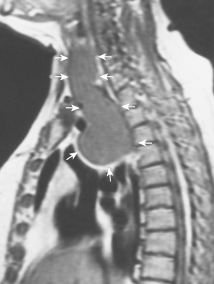
Dermoid cysts and teratomas have similar imaging features. They typically appear as heterogeneous, sharply marginated, multiloculated, cystic, anterior mediastinal masses. A combination of fluid, fat, and calcification is frequently observed. This unique combination of findings makes teratoma one of the few mediastinal tumors that can confidently be diagnosed by radiographic findings alone. A fat-fluid level is seen in 10% of cases and is highly specific for teratoma (Fig 16-8). The identification of dental tissue, such as a well-formed tooth, is rare, but it is also diagnostic of this entity. If a dominant, solid soft tissue component is observed within the mass, a malignant GCN or a teratoma with malignant components should be considered in the diagnosis.

Figure 16-8 Teratoma. A, The lateral chest radiograph shows increased opacity in the normally clear retrosternal space, with a well-defined, round border inferiorly (arrow). B, Axial, contrast-enhanced CT image reveals a round, anterior mediastinal mass with a partially calcified rim (right arrow) and a fat-fluid level (down arrow). The latter finding is essentially diagnostic of a teratoma.
Surgical excision of teratomas and dermoid cysts is usually curative. Thorough pathologic sampling is recommended to exclude small foci of immature tissue, other germ cell tumors, or carcinoma. In contrast, patients with malignant GCNs usually have a poor prognosis, with the exception of those with seminoma. Mediastinal seminomas are usually radiosensitive, and patients have an overall survival rate of about 75%.
Thyroid Abnormalities
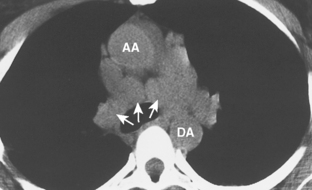
Thyroid abnormalities account for most thoracic inlet masses in adults (Box 16-6). They may extend inferiorly into the anterior, middle, and posterior compartments of the mediastinum. When located in the anterior mediastinum, thyroid masses are almost always located posterior to the great vessels, usually in a paratracheal location. However, most other anterior mediastinal masses are located anterior to the great vessels, and a mediastinal mass located anterior to the great vessels in a retrosternal location is unlikely to be of thyroid origin.
Most mediastinal masses of thyroid origin represent thyroid goiters, and they almost always extend inferiorly from the thyroid gland. A truly ectopic thyroid goiter is rare. Other thyroid abnormalities, such as thyroid adenomas and malignant thyroid neoplasms, infrequently extend into the mediastinum.
Because thyroid goiters account for most mediastinal masses of thyroid origin, the demographics of thyroid mediastinal masses are similar to those of thyroid goiter, with a tendency to occur predominately in middle-aged women. Most patients are asymptomatic, but symptoms may arise from compression of the trachea or esophagus.
CT imaging features of mediastinal thyroid goiters include continuity of the mass with the cervical thyroid gland; foci of high attenuation on non–contrast-enhanced examination (reflecting high iodine content of thyroid tissue); foci of heterogeneous attenuation (i.e., low attenuation cystic areas and high-attenuation foci of calcification); and intense and prolonged enhancement after administration of intravenous contrast. As on plain radiographs, deviation or compression of the trachea is frequently identified on CT. Large thyroid masses, especially posterior descending goiters, may compress the esophagus.
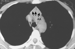
The most important of these features is demonstration of continuity of the mass with the cervical thyroid gland. A combined CT examination of the lower neck and chest is best (Fig. 16-9), although MRI can be used (Fig. 16-10). A radioiodine scan may be confirmatory, with demonstration of radioiodine uptake from foci of functioning thyroid tissue within the mass.

Figure 16-9 Thyroid goiter. A, The posteroanterior chest radiograph shows a large, anterior mediastinal mass. The superior extent of the mass (upper white arrow) extends above the thoracic inlet and is associated with rightward deviation of the trachea (black arrows). The mass extends inferiorly to the level of the base of the heart (lower white arrows). B, Axial, contrast-enhanced CT image of the lower neck shows a heterogeneous mass (open arrows) that is continuous with the isthmus and left lobe of the thyroid gland. The mass contains foci of thyroid tissue that demonstrate intense enhancement and foci of low attenuation consistent with cysts. C, Axial, contrast-enhanced CT image at the level of the aortic arch shows the large, substernal component of the mass (arrows), which displaces the ascending aorta (A) and superior vena cava (S) posteriorly.
MIDDLE MEDIASTINAL MASSES
Lymphadenopathy
Neoplastic, inflammatory, or infectious lymphadenopathy (Box 16-7) is the most common cause of a middle mediastinal mass (Box 16-8). It is therefore no surprise that there are no distinguishing demographic features.
Box 16-8 Mediastinal Lymphadenopathy Differential Diagnosis
INFECTIOUS CAUSES
Fungal infection (especially histoplasmosis)
Lymphadenopathy should be considered in assessing a middle mediastinal mass when the mass is localized to a known anatomic lymph node site, such as the azygous, subcarinal, or aortopulmonary window regions. Lymphadenopathy often manifests as multiple, discrete masses (Fig. 16-11), in contrast to most other causes of mediastinal masses, which usually manifest as a single mass. Multiple masses within known anatomic lymph node sites suggest lymphadenopathy.

Figure 16-11 Sarcoidosis. Axial, non–contrast-enhanced CT image shows discrete, round, soft tissue masses (arrows) in the anterior subcarinal region, consistent with enlarged lymph nodes. Other images demonstrated lymphadenopathy in the right paratracheal region and both hila, a characteristic distribution of lymphadenopathy in sarcoidosis. AA, ascending aorta; DA, descending aorta.
CT plays a role in detecting and characterizing lymph nodes. Although lymph nodes often appear as homogeneous soft tissue density on CT, they may also demonstrate calcification, low-density centers, or vascular enhancement. Identification of these lymph node characteristics can shorten the lengthy differential diagnosis of mediastinal lymphadenopathy (Box 16-9).

Full access? Get Clinical Tree

