Mediastinum


Mediastinum


Paul G. Thacker

Kushaljit S. Sodhi

I. Nimala A. Gooneratne

Claudio Fonda

Pierluigi Ciet

Edward Y. Lee


INTRODUCTION

The mediastinum represents the intrathoracic compartment bounded by the sternum anteriorly, the vertebral column posteriorly, the parietal pleura laterally, the thoracic inlet superiorly, and the diaphragm inferiorly. It is the most common location of chest masses in the pediatric population.1,2 In addition to various normal variants, a wide spectrum of disorders affects the mediastinum including congenital vascular and nonvascular anomalies, infectious and inflammatory diseases, and benign and malignant neoplastic lesions.1,2 Furthermore, traumatic injury may also affect mediastinal structures in pediatric patients.

It is, thus, imperative for the radiologist to have an up-to-date knowledge of the various imaging techniques currently available for optimal evaluation of both normal anatomy and pathologic mediastinal processes. In addition, the radiologist should be aware of characteristic imaging appearances of this broad spectrum of normal variants as well as congenital and acquired abnormalities. This chapter presents a comprehensive review of the pediatric mediastinum including currently available imaging techniques, the normal mediastinal anatomy, and the broad spectrum of mediastinal disorders that occur in infants and children.


IMAGING TECHNIQUES

Understanding the various modalities available for imaging the mediastinum, their strengths and weaknesses, as well as the most appropriate modality for assessing various mediastinal entities is of paramount importance for developing an accurate and cost-effective diagnostic imaging plan. In general, there are four primary goals for imaging mediastinal abnormalities: (1) identifying mediastinal pathology, (2) characterizing identified pathology, (3) providing a succinct and accurate differential diagnosis, and (4) generating a cost-effective strategy for additional imaging and patient management. All of the radiology modalities currently available, that is, radiography, ultrasound, computed tomography (CT), magnetic resonance imaging (MRI), and nuclear medicine imaging, have utility for the evaluation of the mediastinum.


Radiography

The first diagnostic modality for the evaluation of the mediastinum in infants and children is generally a frontal and lateral chest radiograph. Relative to other modalities, chest radiography has a widespread availability, low cost, and ease of acquisition. However, one disadvantage that should be considered is the ionizing radiation associated with chest radiographs, particularly in the pediatric population who are vulnerable to potentially harmful ionizing radiation. Exact technique varies depending on the patient age and size, but fields of view of chest radiographs generally extend from the base of the neck to the proximal upper abdomen. In the very young and the very sick, only a supine frontal radiograph may be obtainable and suffice. Occasionally, a cross-table lateral radiograph may be added in these situations if necessary. Nevertheless, if feasible, upright frontal and lateral radiographs should be obtained for optimal assessment and characterization of mediastinum in pediatric patients.


Ultrasound

With respect to the mediastinum, the role of ultrasound is limited in older children who are generally older than 5 years because of a suboptimal acoustic window. Conversely, in the
young child and infant, ultrasound can be particularly helpful and may be the next imaging modality of choice after an initial radiograph. Advantages of ultrasound include its widespread availability, portability, capability for real-time evaluation of the mediastinum, and its lack of ionizing radiation.

The optimal transducer used for evaluation of the mediastinum varies with age. In neonates and infants, 5- to 10-MHz linear array transducers are generally used whereas older children and adolescents may require 2- to 4- or 4- to 7-MHz sector or linear array transducers for adequate tissue penetration.3 Depending on the age of the child and the location of the process, suprasternal, sternal, parasternal, intercostal, or subxiphoid transducer positions may be used.3 In a young child, the unos-sified sternal and costal cartilage provides an adequate acoustic window for the evaluation of the mediastinum.1 Imaging is generally performed in the supine position, but additional prone and decubitus positions may be of value. Doppler interrogation may be helpful for the complete evaluation and characterization of both solid and cystic mediastinal lesion.


Computed Tomography

Because of its high spatial resolution and capacity for multiplanar reformations and three-dimensional reconstruction, CT plays a pivotal role in the evaluation of the pediatric mediastinum, particularly in the setting of mediastinal masses. CT has been shown to have a very high diagnostic accuracy for the characterization of mediastinal masses in terms of size, location, nature, and additional organs of involvement.1,4 Furthermore, outside of the primary lesion, CT has been shown to provide additional diagnostic information in 82% and affected clinical management in 65%.1,4,5

Typical CT parameters for mediastinal evaluation should closely adhere to the ALARA (As Low As Reasonably Achievable) principle by using the lowest possible radiation dose to achieve diagnostic value. Given the variance in patient sizes for different age groups in the pediatric protocol, CT parameters can be either weight based or girth based. Thin collimation (<1 mm) and fast table speeds of <1 second are available for use on most modern multidetector computed tomography (MDCT) scanners. In evaluation of mediastinal masses in the pediatric population, obtaining noncontrast CT images is not necessary because it does not typically provide additional information. Intravenous contrast is useful particularly in pediatric patients given the lack of mediastinal fat and the advantage over noncontrast images for the full assessment of mediastinal masses. If at all possible, power injection technique, which provides more homogeneous contrast enhancement, is preferred. However, this often depends on the type, location and stability of venous access available. Postprocessing techniques such as 2D multiplanar reformations and 3D reconstructions have been shown to be beneficial in the characterization of mediastinal vessels, central airway anomalies, and mediastinal mass associated abnormalities.


Magnetic Resonance Imaging

MRI is uniquely suited for the evaluation of the mediastinum given its high tissue contrast resolution relative to other techniques and its lack of ionizing radiation. However, it has several important disadvantages that deserve consideration including high cost, susceptibility to motion artifact, need for sedation in young children, and limited spatial resolution of the lungs.1,6,7,8,9,10,11,12,13,14 MRI is particularly advantageous for the evaluation of thymic neoplasms with adjacent mediastinal structure and chest wall involvement, characterization of thoracic lymphatic malformations (LMs), foregut duplication cysts with high proteinaceous content, and intraspinal extent in neurogenic tumors.1

MRI protocols for the evaluation of mediastinal lesions vary somewhat based on the scanner type and institutional preferences. However, some general principles and sequences should be employed. Commonly, an eight-channel cardiac coil is recommended for mediastinal evaluation if available. Sedation is often required in younger patients (<6 to 8 years old) with the exception of neonates who can often be swaddled. Specific MR sequences that are useful include axial fast recovery fast spinecho (FRFSE) T2-weighted imaging with fat saturation; coronal FRFSE T2-weighted imaging with fat saturation; axial T1 or double inversion recovery sequences; coronal gadolinium-enhanced 3D MR angiography spoiled gradient-recalled echo sequences; and axial and coronal postgadolinium T1-weighted imaging with fat-saturation. For the FRFSE T2-weighted MR sequences, a breath-hold or respiratory triggering is recommended to reduce motion artifact. Likewise, ECG gating and a breath-hold are needed for optimal, diagnostic quality double inversion recovery sequences.


Nuclear Medicine

Nuclear medicine imaging of the pediatric mediastinum is primarily limited to positron emission tomography (PET) often co-registered with CT (PET-CT) and metaiodobenzy-guanidine (MIBG) imaging with gallium-67 imaging in lymphoma, primarily of historical interest.

Although not currently a first-line modality for the evaluation of all mediastinal masses, PET-CT has become a near first-line modality for staging, response to treatment, and postcompletion therapy assessment in lymphoma patients in recent years.1,15,16,17,18,19,20,21,22,23,24 Specifically, PET is superior to other imaging techniques for the discrimination of viable tumor from scar and necrotic/nonviable residual tissue, the detection of tumor within normal-sized lymph nodes, and the presence of disease within extranodal sites.1 Typically, the patient is asked to fast for at least 6 hours prior to the administration of 2-fluoro-2-deoxy (18 fluorine)-D-glucose (18FDG). Depending on the Institution, intravenous and oral contrast may be administered. Additionally, co-registered CT technique may vary from full, diagnostic quality technique to very-low-radiation technique, which simply functions for anatomic localization. Although primarily used in the setting of lymphoma, PET-CT may one day find uses in the mediastinum for diseases other than lymphoma, particularly in malignant or metastatic tumors.

A guanethidine analog similar to norepinephrine, MIBG, when tagged to the radioisotope iodine-123, is taken up by chromaffin cells and helpful in the imaging of abnormal sympathetic adrenergic tissue. For the pediatric population, MIBG is mainly employed for the evaluation of suspected or known
sympathetic chain tumors such as neuroblastoma. MIBG imaging has a high detection rate (> 90%) for neuroblastoma. The patient is typically given a dose of 3 to 10 mCi of radioiodinated MIBG radiopharmaceutical. Whole-body planar images are performed 24 to 48 hours after administration.25


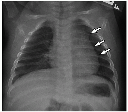
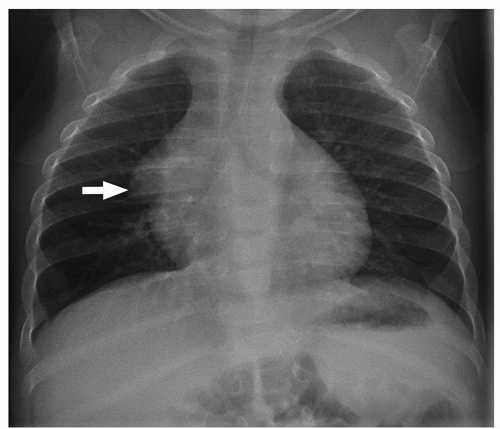
FIGURE 11.1 A 2-month-old girl with normal thymus on frontal chest radiograph. Frontal chest radiograph demonstrates an undulating contour of the left lateral thymic border (arrows). This represents the normal thymus interleaving into the adjacent rib interspaces giving the so-called “thymic wave” sign and should not be mistaken for a mass.


NORMAL ANATOMY AND VARIATIONS


Thymus

The thymus is a bilobed encapsulated organ located anterior to the pericardium and great vessels. It serves as an important immune system organ with its primary function being the maturation of T lymphocytes. Its appearance varies as the patient ages, which can lead the unwary radiologist to mistake the normal thymus for an abnormal mass. This is particularly troublesome in pediatric patients with lymphoma who are receiving chemotherapy because the thymus often involutes while on therapy with subsequent thymic rebound simulating tumor recurrence at the cessation of therapy. Furthermore, ectopic thymic tissue and thymic variants may also introduce diagnostic dilemma (Figs. 11.1 and 11.2). The two most common thymic variants are superior extension of the thymus to the level of the lower neck (Fig. 11.3) and posterior extension
of the thymus generally posterior to the superior vena cava or posterior to the aortic arch (Fig. 11.4). Recognition of the connection between the ectopically located portion of the thymus and normally positioned portion of the thymus as well as the lack of mass effect upon adjacent mediastinal structures are two helpful clues to confirm the diagnosis of ectopically located but normal thymus.


FIGURE 11.2 A 3-month-old boy with normal thymus on frontal chest radiograph. Frontal chest radiograph demonstrates the “thymic sail” sign representing the lateral triangular extension (arrow) of the normal thymus. This should not be confused for the “spinnaker sail” sign (Fig. 11.50) whereby the thymus is uplifted by pneumomediastinum giving the contour of a spinnaker sail.


FIGURE 11.3 A 5-year-old boy who underwent chest MRI for chest wall asymmetry. A: Axial T1-weighted MR image shows a soft tissue, mass-like structure (arrow) located at the level of thoracic inlet. B: Sagittal postcontrast T1-weighted MR image demonstrates an ectopically located portion (asterisk) of the thymus, which is connected (arrow) to the normally positioned thymus (T), consistent with superior extension of the thymus.


FIGURE 11.4 A 3-month-old boy who underwent MRI examination for possible mediastinal mass. Axial T2-weighted MR image shows posterior extension (arrow) of the normal thymus.

On chest radiography, the appearance of the thymus varies by patient age (Fig. 11.5). During infancy, the thymus is typically quadrilateral in shape with convex outer margins. After around age 5, the thymus becomes more triangular in shape with straightening of its margins. After approximately age 15, the margins of the triangular-shaped thymus begin to become more convex with the thymus slowly involuting over time into adulthood. Any outward, particularly mass-like, convexity of the margins after approximately age 5 should raise the suspicion for an abnormal thymic mass. However, thymic rebound related to the relief of certain stressors such as the cessation of chemotherapy, recent surgery, or intubation must also be considered (Fig. 11.6).

If question remains after review of the initial chest radiograph, ultrasound may be utilized in children <5 years of age to confirm the presence of a normal thymus. On ultrasound, the normal thymus appears as a smoothly marginated, sharply defined anterior mediastinal organ, which closely molds to the underlying structures (Fig. 11.7). The pliable thymus typically deforms with cardiac and vascular pulsations. There is uniform echogenicity throughout the thymus, which most closely resembles that of the liver with hyperechoic septations interspersed throughout the gland.


FIGURE 11.5 Three axial CT images of a normal thymus in patients aged 6 months (A), 5 years (B), and 17 years (C).

Generally, the normal thymus does not require CT or MRI for evaluation. However, the normal thymus is quite frequently demonstrated on these modalities when imaging is performed for alternative indications. Therefore, it is important to understand the CT and MRI appearance of the normal thymus. The macrostructure of the thymus on CT and MRI correlates closely with that on ultrasound, that is, smoothly marginated, homogeneous glandular tissue, which conforms to the adjacent structures. There should be no associated compression or displacement of the surrounding anatomy. On CT, the normal thymus shows a homogeneous attenuation value similar to that of chest wall musculature (Fig. 11.5). On MRI, the signal intensity of the normal thymus is typically slightly higher than that of adjacent thoracic muscle on T1-weighted MR images (Fig. 11.3) and slightly less than or equal to that of fat on T2-weighted and fat-saturated MR images (Fig. 11.4).


Lymph Nodes

Lymph nodes are oval, bean-shaped, or rounded soft tissue structures located along the course of lymphatic chains and consist of a fibrous capsule with multiple internal trabeculae, which help to support and contain lymphatic tissue. They are present throughout the mediastinum. For accurate localization, lymph nodes have been regionally classified by the American Thoracic Society into four nodal regions, that is, superior mediastinal nodes, aortic nodes, inferior mediastinal nodes, and N1 nodes, with a total of 14 nodal stations.26

Unlike in adults, there are currently no established size criteria for normal mediastinal lymph nodes in children.27 As a general rule, normal mediastinal lymph nodes should not be viewable on imaging, particularly chest radiography, prior to puberty. However, as more and more children are receiving advanced cross-sectional imaging with ever improving spatial resolution, it is conceivable that tiny mediastinal lymph nodes become viewable in the absence of disease. However, at present, any mediastinal lymph nodes seen on imaging should be noted, and any underlying causes of lymphadenopathy should be excluded.28


Azygoesophageal Recess

The azygoesophageal recess (AER) is a mediastinal space representing protrusion of the medial border of the right lower lobe into the mediastinum from the level of the azygos arch inferiorly to the level of the aortic hiatus and right hemidiaphragm.29


FIGURE 11.6 Thymic rebound in a 10-year-old girl status post chemotherapy. A: Axial enhanced CT image through the upper chest demonstrates a quadrilateral-shaped, smooth, and homogeneous thymus consistent with thymic rebound. B: Axial enhanced follow-up CT image in the same patient through the thymus demonstrates return of normal triangular thymic shape and size.

On frontal chest radiographs, the AER appears as a vertically oriented interface projected over the thoracic spine. On cross-sectional imaging, the AER appears as a focus of aerated lung protruding and extending across the spine a variable distance and bordered anteriorly and medially by the esophagus, the left atrium, and the azygos vein (Fig. 11.8). In adults, it is most often convex to the left (dextroconcave).29 However, children demonstrate a more varied appearance ranging from dextroconvex, to straight, to dextroconcave. Multiple abnormalities affect the AER including developmental anomalies such as foregut duplication cysts, esophageal abnormalities particularly inflammation, vascular anomalies, lymphadenopathy, and neurogenic tumors.29


Mediastinal Compartments

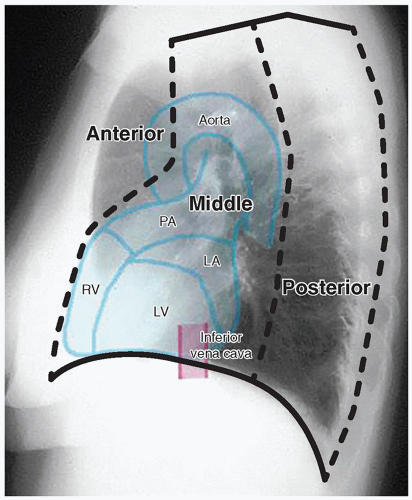
The mediastinum compartmental division is classically based on the lateral chest radiograph and artificially divided into three compartments, that is, anterior, middle, and posterior (Fig. 11.9). These compartments are important because localizing mediastinal pathology to a particular compartment is often the first step in imaging evaluation. It also substantially helps in limiting the differential diagnosis and aiding in the decision of which additional imaging modality or modalities need(s) to be subsequently obtained in reaching the definitive diagnosis.1


FIGURE 11.7 Normal thymus in a 6-week-old girl. The normal thymus (arrows) is located anterior to the mediastinal vessels and demonstrates a homogeneous appearance with linear and punctate echogenic foci. No mass effect upon adjacent aorta (A) or superior vena cava (SVC) is seen.

Recently, the International Thymic Malignancy Interest Group (ITMIG) developed and published a three compartmental mediastinal classification system based on axial CT anatomic divisions.30 This three compartmental system includes the prevascular compartment, the visceral compartment, and the paravertebral compartment. These three compartments are similar to the radiographically based anterior, middle, and
posterior compartment. For the purposes of this chapter, we refer to the classic classification system, as, for our purpose, the compartmental classifications are roughly interchangeable.


FIGURE 11.8 Azygoesophageal recess (AER) in two different pediatric patients. A: Axial enhanced CT image from a 4-year-old girl demonstrates the AER (arrow) with a convex lateral shape, resulting from intrusion of the esophagus (E) into the AER. B: Axial enhanced CT image from a 11-year-old girl shows the AER (arrow) with a straight border.


FIGURE 11.9 Lateral radiograph with three mediastinal compartments. Anterior, anterior mediastinal compartment; Middle, middle mediastinal compartment; Posterior, posterior mediastinal compartment.

The anterior mediastinal compartment is a lenticular space bordered by the sternum anteriorly and the pericardium posteriorly. Often times, it is helpful to consider which normal structures lay within a particular mediastinal compartment because this can aid in formulating the most likely differential diagnosis. Normal structures within the anterior mediastinal compartment include the thymus and lymphatic tissue. The middle mediastinum constitutes the largest mediastinal compartment. It lies between the anterior pericardial border and an imaginary line drawn ˜1 cm posterior to the anterior border of the vertebral bodies. The superior border is formed by the thoracic inlet, whereas the diaphragm represents the inferior border. Normal structures within the middle mediastinal compartment include the heart, great vessels, trachea, esophagus, and lymph nodes. The posterior mediastinal compartment is anteriorly bordered by an imaginary line drawn ˜1 cm posterior to the anterior border of the vertebral bodies, posteriorly by the posterior paravertebral gutters, superiorly by the thoracic inlet, and inferiorly by the posteromedial slips of the diaphragm. The normal structures of the posterior mediastinal compartment consist predominately of parasympathetic and sympathetic nerve chains and the osseous structures of the vertebral column (Table 11.1).


TABLE 11.1 Spectrum of Non-vascular Mediastinal Masses in the Pediatric Population


Anterior Mediastinum


Middle Mediastinum


Posterior Mediastinum


Thymic hyperplasia


Congenital foregut duplication cyst


  • Bronchogenic cyst


  • Esophageal duplication cyst


  • Neurenteric cyst


Neuroblastic tumor


  • Neuroblastoma


  • Ganglioneuroblastoma


  • Ganglioneuroma


Lymphoma


  • Hodgkin lymphoma


  • Non-Hodgkin lymphoma


Lymphadenopathy


  • Infectious lymphadenopathy


  • Neoplastic lymphadenopathy


  • Castleman disease


Peripheral nerve sheath tumor


  • Neurofibroma


  • Schwannoma


Thymic cyst


Thymoma


  • Invasive type


  • Noninvasive type


Thymic carcinoma


Germ cell tumor


Thymolipoma and lipoma


Lymphatic malformation


SPECTRUM OF MEDIASTINAL DISORDERS


Anterior Mediastinal Lesions


Thymic Hyperplasia

Also known as thymic rebound, thymic hyperplasia represents enlargement of the thymus after a period of atrophy induced by medications or severe illness. In the setting of chemotherapy, the thymus atrophies in 90% of patients.2 After the illness has subsided or the medication administration has ceased, the thymus slowly recovers in volume with the total size possibly exceeding its baseline value. Imaging clearly demonstrates this pattern of involution and subsequent regrowth.


FIGURE 11.10 Hodgkin lymphoma of the mediastinum in a 13-year-old girl, forming a mass with a fleshy cut surface (left). Microscopically, large atypical cells (“Reed-Sternberg cells”) are present within a background of mixed inflammatory cells (right; hematoxylin and eosin; original magnification, 600×).

On chest radiographs, the thymic shadow significantly diminishes in size or completely disappears during the associated stressor. Once the illness or medication stress has subsided, the thymic shadow rebounds and may demonstrate lobulated and convex contours, which can be disconcerting for the imager. Similarly, cross-section imaging demonstrates thymic hyperplasia to best advantage (Fig. 11.6). The hyperplastic thymus typically demonstrates homogeneous soft tissue attenuation and signal on CT and MRI, respectively, helping to differentiate it from more ominous processes. On PET imaging, thymic rebound is quite frequently encountered and should be distinguishable from persistent or recurrent neoplasm. In addition to uniform soft tissue attenuation on co-registered CT, homogeneous rather than masslike focal uptake is a supporting finding for thymic rebound on PET. Additionally, the rebounding thymus typically should not have a standardized uptake value (SUV) of more than 4.2

No specific treatment and follow-up is needed for thymic hyperplasia other than distinguishing it from more ominous diseases.


Lymphoma (Hodgkin and Non-Hodgkin Lymphoma)

Lymphoma represents the most common anterior mediastinal mass in childhood, arising from abnormal proliferation of lymphocytes.1,2 Lymphoma is the third most common malignancy in children with only leukemia and central nervous system tumors being more common. Traditionally, lymphoma is subdivided into Hodgkin, histologically characterized by Reed-Sternberg cells and variants (Fig. 11.10), and non-Hodgkin lymphoma (NHL), resulting from clonal proliferation of lymphocytes (Table 11.2).


TABLE 11.2 Lymphomas Occurring in the Pediatric Mediastinum


T lymphoblastic leukemia/lymphoma


Classical Hodgkin lymphoma


Primary mediastinal large B-cell lymphoma


Anaplastic large cell lymphoma


B-cell lymphoma, unclassifiable, with features intermediate between diffuse large B-cell lymphoma and classical Hodgkin lymphoma


Other mature B-cell lymphoma


Hodgkin lymphoma in children typically occurs after the age of 10 years. It more often affects boys than girls by a ratio of 2:1.31 It is divided into classical Hodgkin lymphoma and nodular lymphocyte-predominant Hodgkin lymphoma. In classical Hodgkin lymphoma, Reed-Sternberg cells and variants are the characteristic malignant cell (Fig. 11.10); they are present in a background rich in benign lymphocytes and other inflammatory cells with variable amounts of fibrosis. Nodular sclerosis subtype, most often showing prominent interspersed fibrous bands, is the most common subtype in the pediatric mediastinum. Nodular lymphocyte-predominant Hodgkin lymphoma is rare in children.32 Generally, Hodgkin lymphoma carries a good prognosis with a cure rate of ˜90%.2,31 The majority of affected pediatric patients present with chest pain and discomfort related to enlarged mediastinal lymph nodes with associated compressive effects on the adjacent airway and vascular structures. However, affected children occasionally present with asymptomatic cervical or axillary lymphadenopathy. Additional signs and symptoms include fever, night sweat, and unexplained weight loss. These constitutional symptoms are known as B symptoms, and they affect the disease staging. Based on the Ann Arbor staging classification, both Hodgkin lymphoma and NHL (discussed below) are categorized into four stages (Table 11.3).33

NHL, which is more common than Hodgkin lymphoma, typically presents in the first and second decades of life but generally occurs in the pediatric patients younger than 5 years of age.1,6,31 Similar to Hodgkin lymphoma, boys are affected more commonly, with a male-to-female ratio of 3:1.31 Four subtypes of NHL are particularly prevalent in children: (1)
lymphoblastic lymphoma (most commonly with a T-cell phenotype) (Fig. 11.11), (2) Burkitt lymphoma, (3) diffuse large B-cell lymphoma (Fig. 11.12), and (4) anaplastic large cell lymphoma. With T-cell lymphoblastic lymphoma, affected pediatric patients commonly present with a mediastinal mass. In contrast, Burkitt lymphoma commonly presents in the abdomen, most frequently around the terminal ileum. Diffuse large B-cell lymphoma presents variably and may involve the mediastinum, often with fibrosis that imparts nodularity.31,34 Symptoms of mediastinal NHL are often related to compression or obstruction of adjacent airway and vascular structures.


TABLE 11.3 Ann Arbor Staging Classification for Hodgkin and Non-Hodgkin Lymphomas


Stage 1


Involvement of a single lymph node region or a single extralymphatic organ


Stage II


Involvement of 2 or more lymph node regions on the same side of the diaphragm or localized involvement of a single extralymphatic organ in association with regional lymph node involvement on the same side of the diaphragm


Stage III


Involvement of lymph node regions on both sides of the diaphragm (III), which may have accompanying extralymphatic extension in association with adjacent lymph node involvement (IIIE) or splenic involvement (IIIS) or both (IIIE, S)


Stage IV


Diffuse involvement affecting one or more tissues or organs outside of the lymphatic system, with or without nearby lymph node involvement


A: Without B symptoms


B: Fever, night sweats, weight loss of >10% body weight over the last 6 months


FIGURE 11.11 Lymphoblastic lymphoma (T-cell phenotype) presenting as a mediastinal mass in a 7-year-old boy. Cells are small, round, and monotonous (hematoxylin and eosin; original magnification, 600×). Flow cytometry (not shown) confirmed a T-cell phenotype.


FIGURE 11.12 Diffuse large B-cell lymphoma, in the mediastinum of a 17-year-old boy. Cells are large with irregular nuclear contours (left; hematoxylin and eosin) and show immunohisto-chemical expression of the B-cell marker CD20 (right; original magnification, 600×).

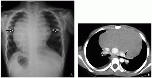
Imaging features of pediatric lymphoma rely heavily on the underlying histology. For Hodgkin lymphoma, chest radiographic findings are variable, ranging from completely normal with minimal mediastinal lymph node enlargement to a very large anterior mediastinal mass (Fig. 11.13A). Following chest radiography, cross-sectional imaging is typically
performed, usually contrast-enhanced chest CT (Fig. 11.13B). Here, the CT serves the purpose of confirming the presence of an anterior mediastinal mass while allowing for the assessment of adjacent nodal involvement, which helps in disease staging (Table 11.3). Imaging appearance of affected nodes ranges from small individually discernible lymph nodes most commonly in the prevascular and pretracheal region to large or conglomerated, lobulated masses that displace adjacent mediastinal structures.2,31 Hodgkin lymphoma demonstrates variable signal characteristics on MRI, although involved lymph nodes usually have hyperintense signal on T2-weighted and intermediate signal on T1-weighted MR images.


FIGURE 11.13 Hodgkin lymphoma in a 7-year-old boy who presented with fever, weight loss, and chest pain. A: Frontal chest radiograph demonstrates a large, smoothly marginated mediastinal mass (arrows). The hilum and spine are clearly visible through the mass localizing it to the anterior mediastinum. B: Axial enhanced CT image demonstrates a homogeneous, well-defined anterior mediastinal mass. The aorta and pulmonary arteries are displaced posteriorly. The left mainstem bronchus (arrow) is elongated, compressed, and narrowed. There is a partially visualized small right pleural effusion.

Over the past several years, PET and PET/CT are transitioning into the forefront for lymphoma imaging as they provide useful physiologic data in addition to traditional anatomic evaluation. Active lymphoma demonstrates marked increased FDG activity (Fig. 11.14) relative to the mediastinal blood pool, whereas fibrosis and necrotic tissue in the region of prior disease demonstrates no appreciable FDG activity.

Unlike HL, the site of involvement of NHL is more variable than that of HL; common sites include the abdomen, thorax, and head and neck. Thoracic involvement occurs in ˜50% of cases.2,31 On chest radiograph, thoracic NHL presents as a large anterior mediastinal mass with convex outer margins and often displacement of the mediastinal structures such as mediastinal vessels and trachea (Fig. 11.15A). Discrete or large conglomerated mediastinal lymphadenopathy may be seen on CT and MRI. With contrast administration, NHL may demonstrate heterogeneity (Fig. 11.15B) or central irregular nonenhancing regions representing focal areas of necrosis. Adjacent hilar and subcarinal lymph node chains are frequently involved. Also, pleural-based masses or effusions may be seen, a feature not commonly seen with Hodgkin lymphoma. Like Hodgkin lymphoma, PET is particularly helpful in the assessment of staging, disease activity, and treatment response. Particularly instructive is PET’s ability to discriminate viable tumor from scar and its ability to detect disease within otherwise normalappearing lymph nodes.


FIGURE 11.14 Initial PET/CT of a 16-year-old girl with newly diagnosed Hodgkin lymphoma. Axial fused PET/CT image demonstrates marked FDG avidity seen best within the anterior mediastinal lymphadenopathy with an SUV value of 13.3.


FIGURE 11.15 Non-Hodgkin lymphoma in a 4-year-old boy who presented with face swelling and respiratory distress. A: Frontal chest radiograph shows a large anterior mediastinal mass (arrows) nearly obscuring the entire left hemithorax. B: Corresponding axial enhanced CT image shows a large, heterogeneous soft tissue mass (M) nearly occupying the entire chest with an epicenter within the anterior mediastinum. The superior vena cava (arrow) is compressed and displaced posteriorly, and the trachea is displaced posteriorly and to the right. There is a partially visualized right pleural effusion.

Treatment for both Hodgkin lymphoma and NHL relies primarily on chemotherapy, sometimes with concurrent radiation therapy. After treatment, coarse calcification at the site of prior disease may develop (Fig. 11.16). Comparatively, the cure rate is higher for Hodgkin lymphoma at 90% with a cure rate among pediatric patients with NHL being 80%.31,35 For NHL, bone marrow transplantation may also be utilized as a treatment option.
Oct 13, 2018 | Posted by in PEDIATRIC IMAGING | Comments Off on Mediastinum

Full access? Get Clinical Tree

Get Clinical Tree app for offline access