19
Melanoma
Eugene C. Lin and Abass Alavi
Positron emission tomography (PET) is most useful in staging high-risk disease at diagnosis and evaluation for recurrent disease if surgery is planned. It has a potential role in assessing response and surveillance for recurrence, but the exact value of PET in these settings is not well defined.
- Initial staging1,2
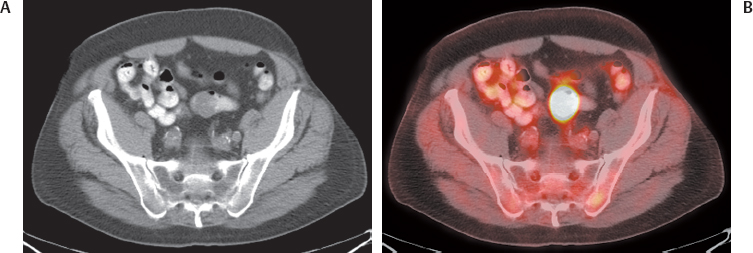
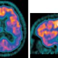
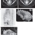
- PET is a useful modality for regional and distant staging of high-risk melanoma at initial diagnosis (Figs. 19.1, 19.2, 19.3, and 19.4).
- The value of PET for initial staging depends upon the disease stage.
- PET is most valuable in stage III disease with regional lymph node metastases.
- Detection of distant disease will likely influence prognosis and management.
- Twenty-two percent of patients will be upstaged by PET.3
- PET has minimal if any role in stage I and II disease.
- In these stages, the prevalence of nodal disease is small, and sentinel node biopsy has a much higher negative predictive value (NPV) than PET. The sensitivity of PET for regional lymph node metastases in melanoma is only 17% relative to sentinel lymphadenectomy.4
- PET may have potential value with a higher pretest likelihood of distant metastases, for example, in melanomas located in the trunk and upper arms, a Breslow thickness > 4 mm, ulceration, or high mitotic rate.4 However, note that in stage I and II disease, possible distant disease identified by PET has a high false-positive rate.5
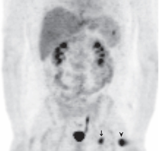
Fig. 19.1 Primary melanoma and metastases. Coronal positron emission tomography scan demonstrates uptake in a primary left thigh melanoma (arrowhead), a left groin nodal metastasis (arrow), and a splenic metastasis.
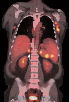
Fig. 19.2 Melanoma metastases. Coronal positron emission tomography/computed tomography scan demonstrates melanoma metastases to the right adrenal (arrow), spleen, and axillary nodes.
- PET may occasionally be of value in stage IV disease.
- PET is usually not helpful if metastases are multiple, as demonstration of additional metastases usually will not alter management. However, PET could be useful in this situation to determine response if experimental therapies are employed.
- In patients with potentially surgically remediable disease (solitary or limited distant metastases), PET is helpful in detecting additional disease, which may obviate plans for surgery.
- PET is more useful in identifying distant disease than regional metastases, given the established role of sentinel node imaging and biopsy. PET and any other gross imaging technique cannot replace sentinel node biopsy because these techniques will not detect micrometastases.6
- However, if used in intermediate- and high-risk lesions, a positive PET study can guide biopsy of appropriate regional nodes.7
- A sentinel node biopsy must be performed if PET is negative, as micrometastases can be missed.
- In general, there is a distinct and appropriate role for both PET and sentinel node biopsy, and either or both should be employed accordingly.
- Recurrent disease1,2
- PET is a standard modality in evaluation of recurrent melanoma. It is useful for
- Diagnosis of recurrent or metastatic disease
- Pretreatment staging of recurrent disease
- The primary indication for PET in recurrence is when surgery is planned.
- Detection of additional sites of disease will avoid unnecessary surgery.
- Equivocal findings on conventional imaging can be characterized.
 Initial Staging and Recurrence