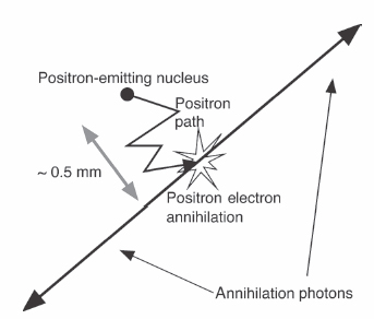
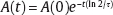
I Positron emission tomography (PET) offers several unique advantages compared with other imaging modalities. PET measures the two annihilation photons that are produced back-to-back after positron emission from a radionuclide tagged tracer molecule, which is chosen to mark a specific function in the body on a biochemical level (Fig. 1.1). Hence, PET provides molecular imaging of biological function instead of anatomy. The detection of both annihilation photons in coincidence yields increased sensitivity over single photon imaging and provides inherent collimation and accurate attenuation correction either from a dedicated transmission scan or from computed tomography (CT) information. This allows extraction of accurate quantitative as well as qualitative information from PET images. Only minute amounts of imaging substrate need to be injected (tracer principle) because of the high sensitivity of PET. In addition, positron emitters (11C, 13N, 15O, 18F, etc.) are relatively short-lived, which enables optimal use of imaging photons while keeping patient radiation dose low. Furthermore, many of these isotopes can be incorporated into biological substrates (glucose, H2O, NH3, CO2, O2, etc.) and pharmaceuticals, without altering their biological activity. Fig. 1.1 General principle of positron emission tomography imaging: decay of radionuclide, positron (β+) emission, multiple scatter in tissue, annihilation with electron, and production of two back-to-back 511 keV annihilation photons. (Not to scale.) Compared with CT scans and magnetic resonance images (MRIs), PET images appear much blurrier or noisier, due to the relatively limited number of photons that can be collected during an imaging study. In addition, detector resolution is poorer due to the detector physics. X-ray CT scanners can easily resolve points < 1 mm in size, whereas PET scanners cannot reliably resolve point sources < 4 to 5 mm at best, and closer to 10 mm in practice. However, this does not impair their high sensitivity to focal tracer concentrations or their usefulness in accurate quantitative functional imaging. In this chapter, we introduce the physics of PET imaging. Several textbooks provide a more in-depth treatment and are included in the References.1,3 Radioactive isotopes are atoms whose inner core, their nucleus, is unstable, in a state with too much energy. Nuclei consist of a densely packed arrangement of protons and neutrons. By undergoing decay, the nuclei change their composition and properties to arrive in a less energetic and more stable state. The decay process follows an exponential law: the number of decays per second is always proportional to the number of undecayed nuclei present. The same is true for the rate of decay, also called activity, which is determined by the half-life of the particular nuclide—the time it takes for half of the original nuclei to decay. Most common in PET is fluorine 18 (18F), which has a half-life of 109 minutes. After some time, t, the activity left, A(t), is proportional to the initial number, A(0), and an exponential term involving the half-life, τ, of the nuclide: Radioactive rates (or activity) are measured in units of becquerel (1 Bq = 1 decay/s) in the International System of Units (SI) or the traditional curie (1 Ci = 3.7 × 10¹⁰ decay/s). A common scale factor used in the clinic is 1 mCi = 37 MBq. In β+ (positron) decay (Fig. 1.1), a nuclide transforms one of its core protons (p) into a neutron (n) and emits a positron (β+), essentially a positively charged electron, and a neutrino (v): p → n + β+ + v. The average positron range in matter depends on the positron’s energy and material characteristics, such as the density and the atomic number. For [fluorine 18]fluorodeoxyglucose ([18F]FDG), positron ranges are rather short, typically < 1 mm. At the end of its path, the positron, being antimatter to electrons, will annihilate (re-combine) with an atomic electron. In the annihilation, electron and positron convert their mass into energy and produce a pair of 511 keV annihilation photons traveling in opposite directions. The 511 keV photon energy (E) comes from Einstein’s famous equation E = mc 2, where m is the mass of the electron or positron (a very small number), and c is the speed of light (a very large number squared). This annihilation radiation is what is detected in PET and what is used to form images of tracer concentration in the body. The dominant annihilation photon interaction in human tissue is Compton scatter. The photon interacts with an electron, ejecting it from its atomic shell. The photon experiences a loss of energy and an associated change of direction, typically out of the detector, and so is unavailable for image formation. Compton scatter and other interactions lead to an attenuation of the annihilation photons. The number of photons that are observed in a straight line from where they were produced decreases exponentially with increasing length of the material traversed. The thickness of soft tissue required to reduce the intensity of a beam by one half is ~7 cm, as opposed to 3 to 4 cm for x-rays. Thus, for ~14 cm of soft tissue, the 511 keV annihilation photon flux would be reduced to one fourth of its original intensity; through the abdomen the photon flux can be reduced to 1/50 of its original intensity. Thus, attenuation is often the dominant factor in PET image quality, especially for thicker patients. The general goal of photon detection is to measure the total energy deposited by the photon when it traverses the detector. For highest sensitivity and accuracy, all of the photon’s energy should be deposited, but in practice this is not always possible. In scintillation crystals, the incident annihilation photon (nominally 511,000 eV energy) interacts and creates tens of thousands of visible wavelength photons (~1 eV energy each) in a very short flash, or “scintillation.” The number of scintillation photons produced in the crystal is proportional to the energy deposited by the annihilation photon. Scintillators for PET photon detection can be rated on four of their characteristic properties: The stopping power is the inverse of the mean distance traveled by photons before they deposit energy in the crystal. This length depends on density and effective atomic number (Z) of the material. A short travel distance is favorable because it will yield more interactions with the 511 keV photons and a better efficiency for detecting them in crystal of fixed size. The decay constant describes how long the scintillation flash lasts in the crystal. Shorter decay constants are desirable because they allow for counting higher photon rates and lower background rates. Good energy resolution—a small ratio of energy variance over energy—means that there are only small fluctuations in the energy measurement. This gives a means to distinguish against PET photons that have Compton scattered (and lost energy) before being measured. The energy resolution depends on the light output and the intrinsic energy resolution of the crystal. The light output, as the name indicates, is the number of scintillation photons produced by each incident photon. Again, this should be as high as possible, allowing the best spatial and energy resolution. The most commonly used PET scintillators are listed in Table 1.1. Other materials are being evaluated (e.g., lanthanum bromide [LaBr]). Manufacturers are divided on the choice of material: currently, BGO (bismuth germinate) is favored by General Electric (GE Healthcare, Chalfont St. Giles, UK), LSO (lutetium oxyorthosilicate) by Siemens (Berlin/Munich, Germany), and GSO (gadolinium orthosilicate) by Philips (Philips Medical Systems, Andover, MA). Time-of-flight PET scanners (TOF-PET) use the scintillator LYSO (lutetium yttrium orthosilicate), which has properties that are very similar to LSO. The most commonly used photodetectors for PET are photomultiplier tubes (PMTs). PMTs are vacuum tubes with a photocathode, which produce electrons from incoming light photons that are accelerated and amplified. The resulting electrical current is proportional to the number of initial scintillation photons and therefore to the energy deposited in the scintillation crystal by the PET photon. By segmenting the scintillator blocks, using many small PMTs, or exploiting the properties of position-sensitive PMTs, the location of the photon detection can be determined. The most commonly used setup today is the block detector (Fig. 1.2). Here, small individual scintillation crystals, a few millimeters in size where they face the patient, are tightly packed into blocks, which are typically coupled to four or more small photomultiplier tubes. To determine the interaction position of the annihilation photon from the spread-out scintillation photon signals, the relative outputs from the PMT signals are compared. The calculated location then determines the crystal element to which the photon is assigned.
Basic Science
1
The Physics of PET/CT Scanners
Ruth E. Schmitz, Adam M. Alessio, and Paul E. Kinahan
 What Makes PET Useful?
 Radioactive Decay
General Principles
Positron Emission and Annihilation
Interaction of Photons with Matter
 Data Acquisition
Photon Detection and Scintillation Detectors
Radiology Key
Fastest Radiology Insight Engine