Mesenteric Abnormalities
Ramesh S. Iyer, MD
LEARNING OBJECTIVES
1. List diagnostic considerations for diffuse and multifocal mesenteric abnormalities.
2. Identify mesenteric adenitis on US and CT.
3. Generate a differential diagnosis for mesenteric cysts.
INTRODUCTION
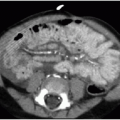
The mesentery is a dual layer of peritoneum that suspends the small and large bowel from the posterior abdominal wall. It is fanor cone-shaped, originating from the superior-central abdomen and radiating inferiorly and peripherally. Mesenteric abnormalities may be divided into focal, multifocal, and diffuse processes. Focal mesenteric lesions are described in the sections below. Diffuse mesenteric pathologies include edema, inflammation, and hemorrhage. These processes typically replace the normal mesenteric fat with soft tissue attenuation on CT, creating a “misty mesentery” appearance, and cause increased echogenicity on US (Fig. 17.1). They also may displace bowel peripherally or encase the superior mesenteric vessels. Multifocal mesenteric masses most often represent lymphadenopathy. Primary diagnostic considerations for mesenteric lymphadenopathy are non-Hodgkin lymphoma, metastatic disease, and infection (Fig. 17.2).1
MESENTERIC ADENITIS
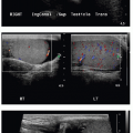
Mesenteric adenitis represents benign nodal enlargement from an underlying viral or bacterial infection. Common bacterial agents include Yersinia enterocolitica and Streptococcus.2,3 In most cases, the terminal ileum is the likely site of infection.4 Presenting signs and symptoms include fever, emesis, leukocytosis, and right lower abdominal pain. Clinically distinguishing mesenteric adenitis from acute appendicitis is challenging, allowing imaging to play an important role in the diagnostic workup.4, 5, 6 and 7
US and CT are the most common modalities for imaging mesenteric adenitis (Figs. 17.3 and 17.4). CT offers a more sensitive and specific evaluation of mesenteric processes, but involves ionizing radiation. There is no clear consensus regarding the normal size of mesenteric lymph nodes in children. Most authors report that a cluster of 3 or more mesenteric lymph nodes with short-axis diameters exceeding 5 mm is required to make this diagnosis
on US (Fig. 17.3).2, 3 and 4,8, 9 and 10 However, others believe that enlarged mesenteric lymph nodes in a child with abdominal pain are a relatively nonspecific finding and that these criteria allow for significant overlap between mesenteric adenitis and normal nodes.9,10 For example, Karmazyn et al.10 reported that using a cluster of 3 or more lymph nodes, each with 8-mm diameters, would be more appropriate to diagnose mesenteric adenitis by CT. The enlarged nodes are most often located in the right abdomen anterior to the psoas muscle.1,4


on US (Fig. 17.3).2, 3 and 4,8, 9 and 10 However, others believe that enlarged mesenteric lymph nodes in a child with abdominal pain are a relatively nonspecific finding and that these criteria allow for significant overlap between mesenteric adenitis and normal nodes.9,10 For example, Karmazyn et al.10 reported that using a cluster of 3 or more lymph nodes, each with 8-mm diameters, would be more appropriate to diagnose mesenteric adenitis by CT. The enlarged nodes are most often located in the right abdomen anterior to the psoas muscle.1,4
Related posts:


Stay updated, free articles. Join our Telegram channel
Full access? Get Clinical Tree
