Mesenteric adenopathy is often much more evident on coronal-reformatted CT
• Ileal ± cecal wall thickening, sometimes with regional ileus
Mucosal hyperenhancement, submucosal edema
• Normal-appearing appendix
TOP DIFFERENTIAL DIAGNOSES
• Appendicitis
• Crohn disease
Early Crohn disease may be impossible to distinguish
Time course and likelihood of recurrence are different
• Cecal or appendiceal carcinoma
Affects older adults, not children
PATHOLOGY
• Reactive lymph node enlargement secondary to enteric pathogens
• Viral (most common)
• Bacterial (especially Yersinia and Campylobacter species)
CLINICAL ISSUES
• Commonly seen in children and young adults < 25 years old
8-12% of young patients with acute RLQ pain have mesenteric adenitis
• Pain, fever, nausea, vomiting
Leukocytosis
• Self-limited, usually resolves without treatment
(Left) Axial CT in a 25-year-old woman presenting with fever and RLQ tenderness shows wall thickening and mucosal hyperenhancement of the terminal ileum and cecum .
(Right) Another CT section in the same patient shows a normal appendix , excluding appendicitis as the diagnosis.
(Left) Coronal-reformatted image in the same patient shows a cluster of mildly enlarged ileocolic mesenteric nodes , along with the thick-walled, inflamed terminal ileum .
(Right) Another coronal CECT section shows enlarged nodes and engorged vessels in the ileocolic mesentery , along with the thick-walled terminal ileum. These are classic imaging and CT features of mesenteric adenitis and enteritis, and the patient made an uneventful recovery without specific therapy.
TERMINOLOGY
Definitions
• Benign inflammation of lymph nodes in ileal mesentery, often with terminal ileitis
IMAGING
General Features
• Best diagnostic clue
Cluster of slightly prominent (≥ 5 mm) mesenteric lymph nodes in right lower quadrant (RLQ)
Ileal/ileocolic wall thickening
Only gold members can continue reading. Log In or Register to continue


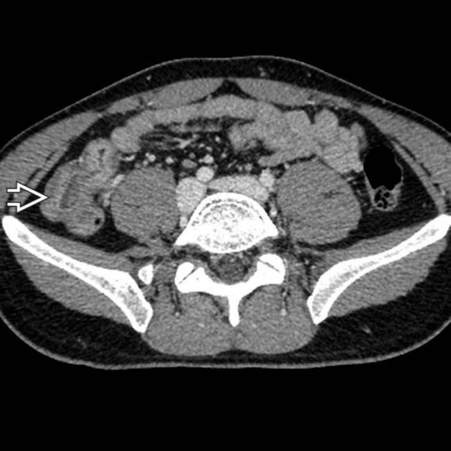
 .
.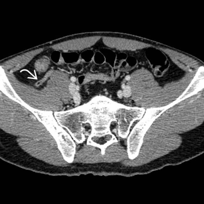
 , excluding appendicitis as the diagnosis.
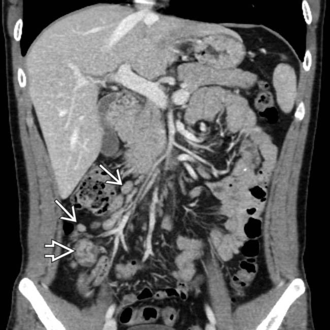
 , along with the thick-walled, inflamed terminal ileum
  .
.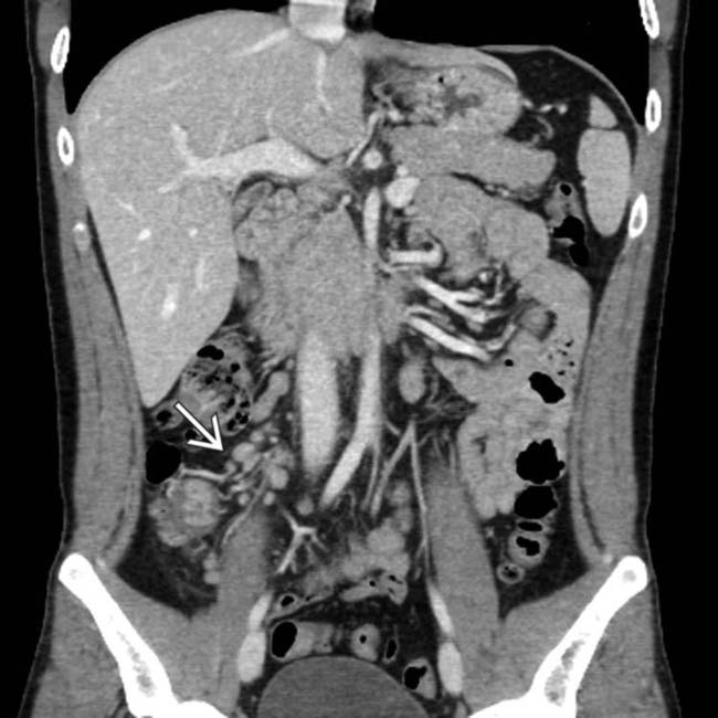
 , along with the thick-walled terminal ileum. These are classic imaging and CT features of mesenteric adenitis and enteritis, and the patient made an uneventful recovery without specific therapy.


