Localization: The most common locations of metastatic carcinoma are the spine, pelvis, femur, ribs, and skull. Metastases may affect any bone but are rare distal to elbow and knee.
Clinical: Pain, mass, pathological fracture, and hypercalcemia. At times (about 20 %) the metastasis to the bone is diagnosed first, without knowledge of the primary lesion even if at the end of staging, primary tumor is often observed in the lung or in the bowel.
Imaging: Standard radiography may be negative (40 % of cases). Frequently, metastases to the bone present as purely osteolytic lesions (kidney), often subperiosteal (lung) or blastic (prostate, breast). Isotope scan and MRI are very sensitive in detecting bone mets (spine!) before radiological changes become apparent on plain x-rays. Today, PET-CT is very useful to detect bone lesions not identified with other techniques and to evaluate relapse of the disease.
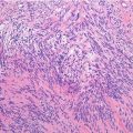
Histopathology: Different features related to the original primary lesion, usually squamous or gland patterns, are present. Origin of metastatic disease is histologically clear only in a few well-differentiated carcinomas (thyroid, kidney, prostate). However, immunohistochemical patterns can help to identify primary tumor.
Treatment: In metastatic patients to maintain or to restore a good quality of life is the most important object. Palliative treatment is necessary when multiple and diffused disease is observed and the patient has a poor prognosis. An adjuvant treatment using chemotherapy, low-dose radiotherapy, and simple surgery is useful in patients with a fair prognosis. In those with a good prognosis, chemotherapy, high-dose radiotherapy, and wide resection with stable reconstruction can be combined together to obtain a long survival. Today, new drugs and new target therapies are more and more spread out in clinical practice.
Key Points
Clinical | More frequent bone lesion. Adults, pain. |
Pathologic fracture | |
Radiological | Permeative or pure lytic, mixed, blastic features |
Histological | Epithelial aspects, depending on primary lesion |
Differential diagnosis | All other primary lesions of adults |
Immunohistochemical Panel
Breast | Prostate | Lung | Thyroid | Kidney | Colon | Urothelial | Melanoma | |
|---|---|---|---|---|---|---|---|---|
CK MNF-116 | + | + | + | + | + | + | + | – |
CK 7 | + | – | + | + | – | – | + | – |
CK 20 | – | – | + | – | – | +
Related posts:Stay updated, free articles. Join our Telegram channel
Full access? Get Clinical Tree
 Get Clinical Tree app for offline access

|