MR imaging of the hip is one of the most common musculoskeletal MR imaging studies performed today to assess for occult fractures, acetabular labral tears, hyaline cartilage loss, and musculotendinous injuries. Several developmental variations are seen in the hip, which can be mistaken for disease or potentially even contribute to the development of a pathologic condition. As in any imaging study, it is important to be cognizant of these variations as well as associated findings that help distinguish between true abnormality and developmental variation when interpreting an MR image of the hip. This article describes the numerous variants of the hip that are frequently seen on arthrographic and nonarthrographic MR imaging examinations.
MR imaging of the hip is one of the most common musculoskeletal MR imaging studies performed today to assess for occult fractures, acetabular labral tears, hyaline cartilage loss, and musculotendinous injuries. Several developmental variations are seen in the hip, which can be mistaken for disease or potentially even contribute to the development of a pathologic condition. As in any imaging study, it is important to be cognizant of these variations as well as associated findings that help distinguish between true abnormality and developmental variation when interpreting an MR image of the hip. This article describes the numerous variants of the hip that are frequently seen on arthrographic and nonarthrographic MR imaging examinations.
Labral anatomy
The acetabular labrum is a fibrocartilaginous horseshoe-shaped structure along the anterior, superior, and posterior margins of the acetabular rim. Unlike the knee menisci, which have a load-bearing and cushioning function, and the glenoid labrum, which helps to deepen the glenoid fossa and improve glenohumeral contiguity, the acetabular labrum primarily seals the margins of the articular hyaline cartilage to prevent premature cartilage loss. However, repetitive impingement and labral tears are a frequent cause of hip pain. Tears occur most commonly in the anterior superior quadrant of the labrum. As a result, evaluation of the labrum to detect abnormalities before the onset of cartilage loss is among the most common indications for imaging of the hip. Occasionally, patients may have asymptomatic labral tears or developmental variations that mimic labral pathology. These patients may benefit from MR arthrography, which distends joint recesses and increases intra-articular pressure, thereby allowing detection of smaller labral tears. Simultaneous intra-articular local anesthetic injection may also be useful to help distinguish between symptomatic and asymptomatic labral tears or developmental variations.
On imaging, the labrum is usually triangular and slightly thicker posterosuperiorly than anteriorly. The labrum usually has a low signal intensity on all routinely used pulse sequences, particularly on more heavily T2-weighted sequences with longer echo times (TE). Several variations in the morphology and imaging appearance of the acetabular labrum can mimic pathologic conditions and lead to ineffective treatment.
Labral tears or detachments can be seen on nonarthrographic MR studies with linear fluid signal tracking into the labral substance or undermining the labral attachment to the acetabular rim. Many tears have intrasubstance high signal on T2-weighted or proton density sequences, with surface irregularity and irregularity of the adjacent hyaline cartilage. Tears can be diagnosed on MR arthrography by visualizing injected contrast material extending into the labrum or through the labrum/acetabulum junction. In more advanced cases, there may be blunting of the labrum, with loss of the normal triangular morphology. Often there is a paralabral cyst with a neck arising either from the labrum itself or from the labro-osseous interface that is commonly seen in association with labral tears.
Intermediate labrum signal
Increased signal within the labrum can be a normal finding, particularly on shorter TE pulse sequences such as T1-weighted or proton density techniques. Hodler and colleagues compared MR images of the hip with histologic findings in cadavers and found that abnormal signal in the acetabular labrum correlated poorly with histologic signs of degeneration. In a study of 52 hips in 46 asymptomatic volunteers, Cotten and colleagues found areas of intermediate or high labral signal intensity in 58% of hips on T1- and proton density–weighted spin echo images in nonarthrographic studies. Intralabral signal was globular, linear, or curvilinear and was located in the superior (87%), posterior (21%), and anterior (8%) labrum. In their study, the abnormal signal was sometimes seen extending to the capsular and/or articular surfaces of the labrum, thus mimicking labral tears in these asymptomatic patients. Although many of these abnormalities resolved on more heavily T2-weighted sequences, intermediate signal persisted in 12% of labra on both T2-weighted conventional spin echo and fast spin echo images. Thus, one should be cautious when relying exclusively on intrasubstance signal alterations to detect labral tears.
Labral shape
The acetabular labrum is normally triangular in cross section, with the lateral part forming the tip and the broader base attached to the bony acetabulum. The labrum is normally thinner anteriorly and thicker superiorly and posteriorly. However, there is a variability in labrum shape in asymptomatic subjects. In their study of MR imaging examinations on 200 asymptomatic hips, Lecouvet and colleagues found the classic triangular labral shape in only 66% of cases, with a round shape in 11% and a flat shape in 9%. They also reported that the prevalence of a triangular labrum decreases with age.
Hypoplasia of labrum
The Buford complex in the shoulder is a well-described normal variant, where there is hypoplasia of the anterosuperior glenoid labrum with an associated enlarged middle glenohumeral ligament. There is debate as to whether a similar phenomenon occurs in the acetabular labrum. Cotten and colleagues reported that the anterosuperior labrum was absent in 10% of asymptomatic hips. Lecouvet and colleagues described an increased incidence of labral nonvisualization with increasing age in asymptomatic patients. In a study of 40 symptomatic patients and 6 cadaveric hip joint specimens using MRI arthrography followed by cryosectioning, Czerny and colleagues found that the labrum was present in all patients and specimens, with no areas of focal absence. Dinauer and colleagues found no instance of an absent portion of the labrum in 58 patients. The finding of absent labral segments may be because of close proximity of the labrum and capsule, precluding resolution of the 2 separate structures. The authors opine that until more work is done that supports focal absence of the labrum is a normal variant, this finding should be considered abnormal.
Cartilage undercutting labrum
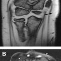
The base of the acetabular labrum normally has a firm attachment to the bony acetabulum. However, on occasion, intermediate signal intensity can be found on MR imaging at the junction of the articular surface of the labrum and the acetabulum. In their study that correlated MR arthrography with anatomic examination of cadaveric hips, Czerny and colleagues showed that this area of increased signal corresponds to the attachment of the labrum to the acetabular cartilage. This should not be mistaken for a tear if the signal is hypointense to fluid or injected arthrographic contrast material, especially if it has a signal intensity similar to that of the hyaline cartilage and smoothly parallels the labral base and acetabular rim. Furthermore, the adjacent hyaline cartilage surface is usually smooth when the findings are related to cartilage undercutting of the labrum ( Fig. 1 ) but may be irregular if there is a true labral tear.
Sublabral sulcus
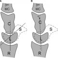
There has been much debate in the literature over the existence and location of a normal sublabral sulcus that could be misinterpreted as a labral tear. In a histologic study of fetal acetabula, Walker and Goldsmith described an anterosuperior sublabral sulcus. However, no definite sulcus was identified on the gross hip specimens, suggesting that the sulcus seen on histology may have been related to a tissue-processing artifact. Dinauer and colleagues described a normal posteroinferior sublabral groove in 22.4% of 58 patients on MR arthrography. In a study on 121 hip arthroscopies, Saddik and colleagues found sublabral sulci in 25% of patients, 44% of which were anterosuperior, 48% of which were posteroinferior, 4% of which were anteroinferior, and 4% of which were posterosuperior. Using a clock face to describe the position of labral findings in which the 12 o’clock position was superior and the nine o’clock position was anterior, Studler and colleagues compared the MR arthrography findings in 57 patients to arthroscopic findings and reported 7 sublabral recesses at the 8-o’clock position, 2 at the 9-o’clock position, and 1 at the 10-o’clock position. They found that a sublabral sulcus does not extend through the entire labral base and that sublabral contrast maintained a linear morphology ( Fig. 2 ). In their study, sublabral sulci showed no abnormal signal in the labrum adjacent to the sulcus, there were no adjacent cartilage lesions or osseous abnormalities, and there were no ganglion cysts. The depth of the labral tears was greater than that of the sulci, but the 2 entities did not differ in longitudinal extent. They concluded that arthrographic contrast material extending through the base of the labrum should be considered as an indication of a labral tear.
Other investigators have disputed the existence of a normal sublabral sulcus. Czerny and colleagues found no evidence of a normal sublabral sulcus in their study of MR imaging arthrograms on 40 symptomatic patients and 6 cadaveric hip joint specimens, and Petersilge and colleagues found no evidence of a normal sublabral sulcus in their study of 10 patients who underwent MR arthrography and subsequent arthroscopy. As a result, many investigators have concluded that there likely is a normal sublabral sulcus posteroinferiorly just above the transverse ligament. However, similar findings in the anterior or anterosuperior labrum should be carefully evaluated because many of these defects represent true labral tears.
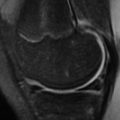
Often imagers rely on direct arthroscopic visualization to determine whether an MR finding represents a tear or a sulcus. However, it should be remembered that arthroscopy is an imperfect gold standard because there is no consensus among arthroscopists about what represents the normal labral appearance. Use of higher-resolution MR imaging, such as 3-tesla imaging, frequently reveals a shallow, smooth defect along the articular surface of the superior and anterosuperior labral base, which may represent a sublabral sulcus ( Fig. 3 ). These defects generally do not involve more than a third of the labral thickness, and the width of the defect is often larger than its depth. In addition, there should not be any adjacent hyaline cartilage loss. On arthrography, contrast should smoothly enter these sulci and should not enter the labral substance.
Transverse ligament
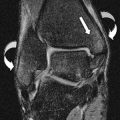
The acetabulum nearly completely covers the femoral head, with the exception of its anteroinferior aspect, where there is a lack of bone and cartilage. This anteroinferior aspect of the acetabulum is spanned by the transverse acetabular ligament, which, with the acetabular labrum, forms a complete ring around the acetabulum. The transverse ligament attaches to the acetabular rim anteriorly and posteriorly and to the ligamentum teres femoris. Where the transverse ligament meets the acetabular labrum, there occurs a normal cleft that can be mistaken for an acetabular labral tear ( Fig. 4 ). Dinauer and colleagues found a cleft at the junction of the transverse ligament and the anterior labrum in 32.8% of hips.
Synovial herniation pits
Femoral fibrocystic changes are common anteriorly at the junction of the head and neck, with some investigators reporting an incidence of up to 33% ( Fig. 5 ). This finding was first described by Pitt and colleagues in 1982. Although the cause is idiopathic, the investigators speculated that the cysts appeared as a result of repetitive herniation of fluid and synovium related to the overlying joint capsule through small perforations in the cortical bone. More recently, Leunig and colleagues proposed that the fibrocystic changes in the anterior part of the bone were not incidental but related to repetitive impingement of the femoral neck and the anterosuperior acetabulum. On radiography, these cysts often appear as lucent lesions in the anterior part of the femoral head/neck junction with a thin peripheral sclerotic rim. They are most commonly small, measuring a few millimeters, but can be larger than a centimeter and may also grow. On MR imaging, the cysts are usually hyperintense to skeletal muscle on T2-weighted sequences and may approach the signal intensity of fluid. Occasionally, there may be marrow edema associated with these cysts, which may manifest as an area of increased radiotracer uptake on bone scintigraphy. A defect in the overlying cortical bone may be seen in larger cysts, and there may be other associated findings of femoroacetabular impingement, such as anterosuperior labral tears, acetabular overcoverage leading to pincer impingement, or femoral head asphericity associated with cam-type impingement. Thus, synovial herniation pits may suggest alterations in hip biomechanics leading to abnormalities, and proper recognition of this finding may help to direct the patient toward proper treatment.