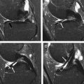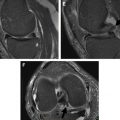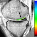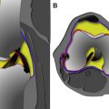
This issue on MR Imaging of the Knee takes a fresh look at the basics of imaging the menisci, ligaments, and tendons; affords a comprehensive look at MR imaging in the pediatric knee; provides new perspectives on the biomechanics of knee injuries and assessment of knee arthritis and regional inflammatory conditions; summarizes the current thinking on MR imaging after surgery on the menisci and on articular cartilage; and addresses the latest and ongoing developments in assessing cartilage degradation as well as MR imaging in the setting of knee hardware. This all-star team of authors has produced a wonderful set of works, and I offer them my heartfelt thanks and gratitude. It is my hope and expectation that this issue will be a useful tool for the radiologist and nonradiologist alike and will provide new, helpful, and exciting information for all those interested in the knee, from trainee to expert. Moreover, I hope that all find this to be enjoyable reading.
Stay updated, free articles. Join our Telegram channel

Full access? Get Clinical Tree


