CHAPTER 2 MRI of the Spine and Bony Pelvis
MRI of the spine and bony pelvis—considerations
• Refer to all safety-related parameters discussed in Chapter 1.
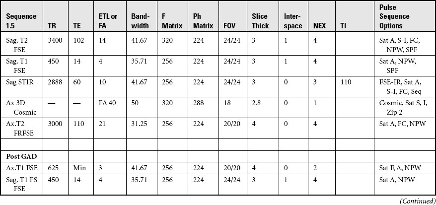
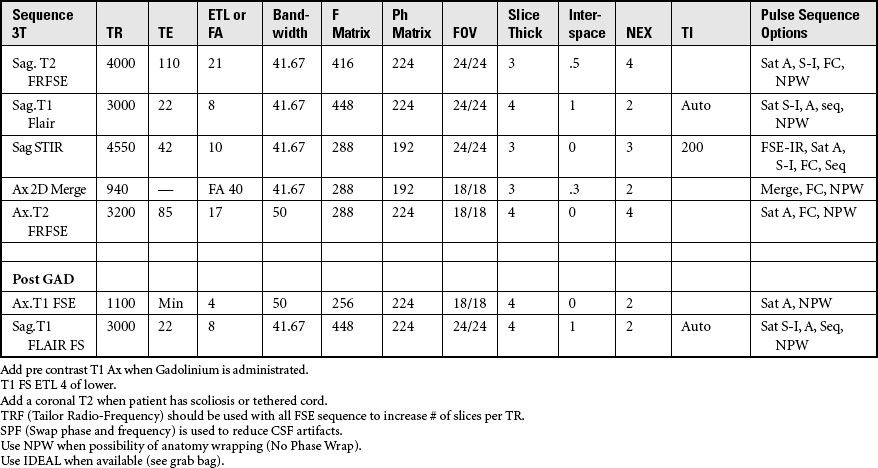
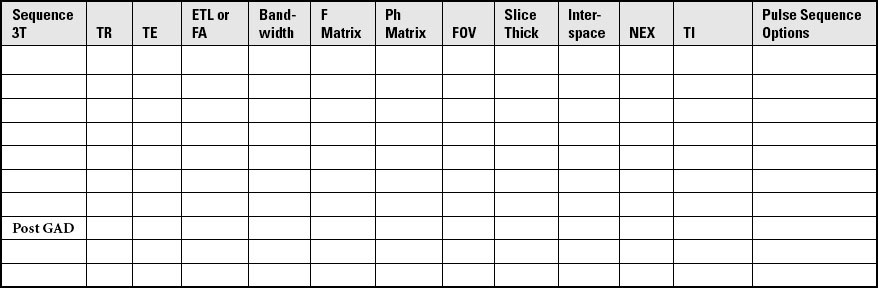
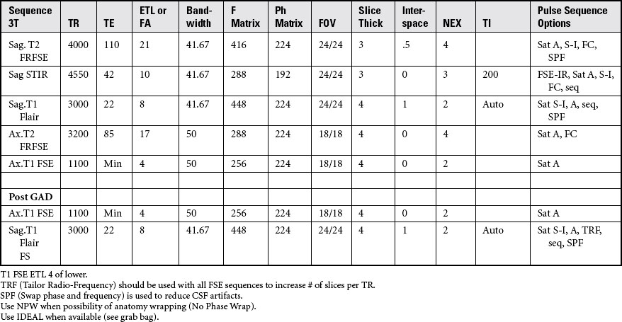
• As the SAR increases, the patient’s body temperature will also increase. At 3 T make sure your estimated SAR is 2.5 or lower before scanning to keep the SAR at an acceptable rate. The SAR elevation can be compensated for by increasing the TR slightly. If this is not done correctly, the SAR will increase and the scanner will stop as a patient safety precaution. All 3-T systems have SAR monitors that should be referenced while scanning. This is not an issue at 1.5 T.
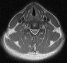
• An eight-channel or higher vendor-specific CTL coil is used primarily to image the spine. The CTL coil is divided into specific sections to correspond to segments of the vertebral column.
• All multichannel coils produce excessive signal adjacent to the coil. This can be compensated for by using vendor-specific options (e.g., GE uses SCIC or PURE to provide uniform signal intensity).
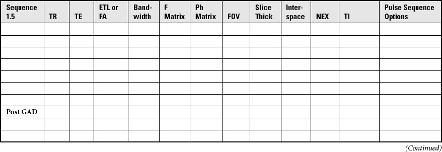
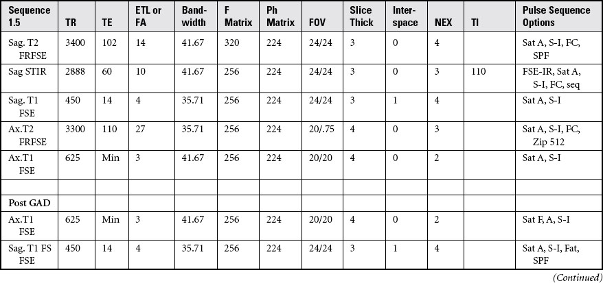
• Generally at 3 T, in the spine, T1 FLAIR is substituted for T1 spin-echo or fast spin echo to compensate for long T1 relaxation times at 3 T.
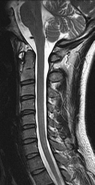
• T1 and T1 FLAIR imaging are best used to identify anatomic structure, whereas T2 and T2 FLAIR imaging provide detailed evidence of pathologic conditions.
• On T1 sequences, CSF produces dark or hypointense signal. T1 FLAIR uses an inversion pulse also to produce dark or hypointense signal.
• On T2 sequences, CSF produces bright or hyperintense signal. T2 FLAIR uses an inversion pulse to produce dark or hypointense CSF signal; all other fluid appears bright.
• Superior and inferior saturation bands help to compensate for CSF and vascular pulsation, whereas anterior saturation bands can help compensate for breathing, swallowing, and peristalsis. Saturation bands can be used on all pulse sequences.
• Fat saturation options and terminology are vendor specific. For GE systems, use “fat classic” for fat saturation with enhanced anatomic detail.
• When trauma, infection, or tumor is suspected, T2 fat saturation or short T1 inversion recovery images should be performed to enhance abnormal fluid or pathology.
• Flow compensation or gradient nulling should be used on T2 images to help compensate for CSF flow and vascular motion. Flow compensation should never be used on T1 precontrast because it causes vessels to appear bright and to mimic pathologic conditions. It is often used post gadolinium on T1 sequences to compensate for flow artifacts.
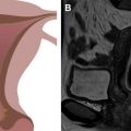
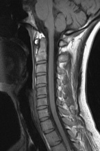
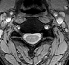
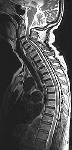
Routine cervical spine
Acquire three-plane pilot per site specifications.
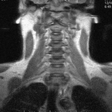
POSITION: Supine, head first, cushion under knees
LANDMARK: Hyoid bone, thyroid cartilage
IMMOBILIZATION: Prevent the body from touching the sides of the magnet by using sponges or sheets.
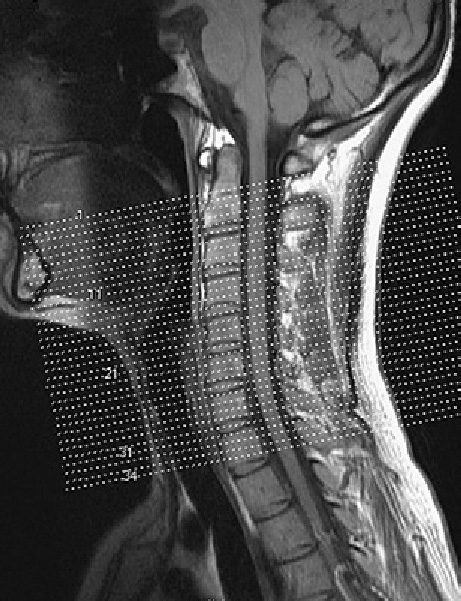
Acquisition of Sagittal Images of the Cervical Spine
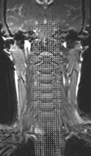
SLICE ACQUISITION: Left to right
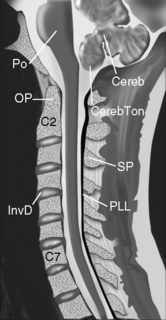
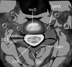
SLICE ALIGNMENT: Parallel to spinal cord and margins of vertebral bodies
ANATOMIC COVERAGE: Cerebellum to thoracic 1, left to right margins of vertebral bodies
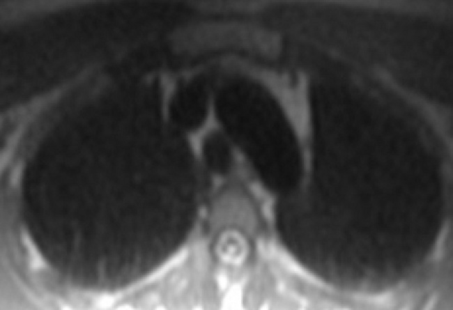
Routine thoracic spine
This allows for accurate identification of vertebral levels.
Acquire three-plane pilot per site specifications.
COIL: Multi-channel or HNS2-3-4
POSITION: Supine, head first, cushion under knees
LANDMARK: Midsternum, 2 to 3 inches superior to xiphoid
IMMOBILIZATION: Prevent the body from touching the sides of the magnet with sponges or sheets. Velcro bands may be used across the torso.