18 MRI Safety As with any procedure, maintaining the safety of the patient and health care team during an MRI examination is of utmost importance, and patient safety considerations begin with the referring clinician. A sound understanding of MRI safety issues not only ensures minimal risk to the patient but also can avoid needless expenses and delays in examinations. Although the MRI center is generally responsible for safety screening, the referring physician can minimize even further the chance of an adverse effect by being aware of important contraindications to MRI and of safety concerns that might put an individual at risk. It can be very comforting to a patient to have the clinician alleviate safety concerns and answer preliminary questions that might otherwise contribute to a growing anxiety in the days leading up to the examination. This chapter reviews these issues and provides guidelines for maximizing safety. In an MRI scanner, the patient’s body is subjected to the baseline static magnetic field and the time-varying magnetic fields created by RF fields and receiver coils. The strength of these magnetic fields is measured in gauss or tesla (T) units; 1 T equals 10,018 gauss. For comparison, the strength of the earth’s magnetic field is 0.6 gauss; the strength of clinical MRI scanners generally ranges from 0.5 to 3.0 T.1 The FDA has guidelines for the exposure of patients to both static and time-varying magnetic fields.2 Currently, the FDA approves clinical imaging using a static magnetic field strength of up to 4.0 T for patients <1 month old and up to 8.0 T for older patients.2 Many studies have evaluated the potential biologic effects of a static magnetic field, and there has been no clear evidence of deleterious effects.3 Although there have been concerns about an elevation of skin and core body temperatures induced by the magnetic field, investigators have concluded that harmful heating does not occur in human subjects.4,5 Reversible electrocardiogram changes (e.g., an increase in the amplitude of the T wave or a nonspecific wave form) secondary to the conductive nature of blood have been noted.6 However, this “magnetohydrodynamic effect” has not been associated with a clinically adverse effect. At strengths of 4 T, patients may experience transient reversible biologic effects, such as nausea (that may be caused by stimulation of the vestibulolabryrinthine complex) or a flashing light sensation (magnetophosphenes, thought to be caused by direct excitation of the optic nerves or retina, by changing magnetic fields, or by rapid eye movements).7 Time-varying magnetic fields or gradient magnetic fields can induce current in the body, which can have two possible biologic effects: heating and neuromuscular stimulation. Even in the absence of metal implants, body temperatures have been shown to rise during MRI, although temperature changes were minor (i.e., <0.6°C).8 The FDA has suggested guidelines to limit the risk of these effects by restricting the strength of time-varying magnetic fields.2 The intensity of the RF energy absorbed by tissue is termed the specific absorption rate and is measured in watts per kilogram. The FDA reports that specific absorption rates of ≤3 W/kg for the head, ≤4 W/kg (averaged) for the rest of the body, and ≤8.0 W/kg for any 1 g of tissue pose no substantial risk.2 Usually, higher specific absorption rate exposures are a concern with faster pulse sequences. Image quality depends on the ability to maintain homogeneity of the magnetic field surrounding the patient. Metallic artifacts can result in misregistered spatial information or signal loss, leading to image distortion. Three types of magnetic materials may cause artifacts because of their inherent abilities to disturb the uniformity of magnetic fields used for imaging: ferromagnetic, paramagnetic, and diamagnetic materials. Of these three, ferromagnetic materials (e.g., iron, nickel, and martensitic stainless steel) concentrate and retain magnetism the most, resulting in severe distortion of the images. Paramagnetic or weakly ferromagnetic materials (e.g., platinum) have a minimal effect on magnetic field homogeneity and result in less image distortion. Diamagnetic materials (e.g., zinc, gold, and copper) do not affect or affect only minimally the static or local magnetic fields. The clinical value of an MRI examination in a patient with metallic hardware depends on the proximity of the hardware to the site of interest. For example, one can anticipate a limited evaluation of neural foramina adjacent to an anterior cervical fusion or an inability to detect an epidural abscess adjacent to pedicle screw instrumentation. Newer pulse sequences have been developed that allow for imaging in the presence of metallic implants (see Chapter 16). Myelography remains a viable alternative for the postoperative spine. CT also is a valuable alternative in postoperative patients, particularly in conjunction with myelography. In addition to causing distortion of images, some metallic foreign bodies or surgical implants can move or generate heat in the presence of a magnetic field, possibly leading to injury or death.9,10 For this reason, each patient must be screened for metallic objects within the body, usually with a verbal interview and a written checklist. Of particular concern are metallic foreign bodies within the orbit that may dislodge and rupture the globe or damage the optic nerve. To our knowledge, there is only one reported case of a patient who suffered a vitreous hemorrhage, caused by a dislodged metal fragment (2.0 × 3.5 mm), that led to blindness.11 Although it is standard for MRI facilities to prescreen for metallic orbital foreign bodies, methods of accomplishing this screening vary. Conventional radiographs of the orbits are said to detect metallic fragments as small as 0.1 × 0.1 × 0.1 mm. However, there is debate over which patients should be referred for conventional radiographs. Many MRI centers obtain orbital radiographs of patients with an occupational history of welding, but some experts advocate obtaining films only if the patient is aware of a previous orbital exposure to metal without removal of the fragment by an ophthalmologist.12 At the authors’ institution, patients with a history of working with metal undergo screening orbital conventional radiographs. Alternatively, review of a previous CT scan, including the entirety of the orbits, is often considered sufficient. In addition to assessing for orbital foreign bodies, the clinician should question the patient regarding any history of metallic foreign bodies near other vital structures, such as the lungs or spinal cord. For example, bullet fragments in the spinal canal contraindicate MRI because they may be ferro-magnetic. The FDA requires testing of all implants to evaluate their safety profiles and classify them as MRI safe, conditionally safe, or unsafe. The list of these devices is extensive and constantly changing, especially as more objects are tested at 3 T and higher, precluding a complete review of these devices in this chapter. A regularly updated list of reviewed implants and devices is available online at http://www.MRIsafety.com. It is important to be aware of devices that absolutely contraindicate MRI scanning, including the following: • Implantable cardiac defibrillators • Cardiac pacemakers • Implantable medication infusion pumps • Ferromagnetic aneurysm clips (e.g., stainless steel) • Poppen-Blaylock carotid artery clamps • Spinal/bone fusion stimulators • Cochlear implants • Tissue expanders • Gastric electrical stimulation devices Certain models of some devices (e.g., implantable cardiac defibrillators, cardiac pacemakers, spinal/bone fusion stimulators, and cochlear implants) may be scanned under strict criteria. However, many MRI centers still consider as absolute contraindications the presence of such devices or of devices that do not meet the required technical conditions (see below). Of note, the Starr-Edwards Model Pre-6018 heart valve prosthesis (Baxter Healthcare, Santa Ana, CA) is now designated as conditionally MRI safe, although previously it was thought to be an absolute contraindication to an MRI examination.13 In recent years, multiple in vitro and in vivo controlled studies have shown increasing evidence that some patients with cardiac pacemakers can be scanned safely under specific conditions, despite the potential for arrhythmia and death.14–18 However, in most situations, a pacemaker remains an absolute contraindication for MRI unless the procedure is an orchestrated effort between cardiology and radiology, with informed consent of the patient.19,20 Similar caution is warranted for patients with neurostimulation systems for deep brain stimulation of the thalamus, globus pallidus, and subthalamic nucleus as a treatment for movement disorders. Although many patients have been scanned successfully without incident, there are specific guidelines regarding the technique.21,22 Currently, the Pulsar Cochlear Implant (MEDEL Corp., Durham, NC) can be scanned, but only under specific conditions on a 0.2-T machine. The Nucleus 24 Auditory Brainstem Implant System (Cochlear Corp., Englewood, CO) has a removable magnet and may be scanned up to 1.5 T, again under specific conditions. It is suggested that the manufacturer of these cochlear devices be contacted before imaging.23 In general, imaging of deep-brain stimulators and this specific implant should be performed only after discussion with a neurologist and radiologist. There are six specific requirements that also must be met to obtain an MRI study of a patient with a spinal/bone fusion stimulator24,25: • Cathodes of the implantable spinal fusion stimulator should be a minimum of 1 cm from nerve roots. • Conventional radiographs should be obtained before MRI to verify that there are no broken leads for the implantable spinal fusion stimulator. • The study should be obtained on an MRI system with static fields of 1.5 T or less, avoiding pulse sequences that expose patient to high levels of RF energy. • The patient should be observed continuously. • To reduce artifact, the implantable spinal fusion stimulator should be placed as far from the spinal canal and bone graft as possible.
 Physiologic Effects of the Magnetic Field
Static Magnetic Fields
Time-Varying Magnetic Fields
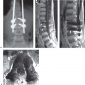
 Metallic Substances
Field Distortion
Metallic Objects in Magnetic Fields
Implant Safety Profiles
Related posts:
Stay updated, free articles. Join our Telegram channel

Full access? Get Clinical Tree


