and Zdeněk Fryšák1
(1)
Department of Internal Medicine III – Nephrology, Rheumatology and Endocrinology, Faculty of Medicine and Dentistry, Palacky University Olomouc and University Hospital Olomouc, Olomouc, Czech Republic
Keywords
Multinodular goiterSolid, complex, or cyst nodulesDominant nodule10.1 Essential Facts
Multinodular goiter (MNG) usually means an enlarged thyroid gland containing multiple thyroid nodules. In adults, the normal thyroid gland has a maximum weight of 18–25 g [1].
In the 1950s, in the era before the ultrasound, prevalence of thyroid nodules at autopsy was: multiple thyroid nodules 37.3%, and solitary nodules 12.2% [2].
At the end of the twentieth century, in the era of ultrasound, prevalence of thyroid nodularity at autopsy ranged from 30–60%, prevalence on palpation was 13–50%, and US imaging reported prevalence 19–67% [3].
The risk of malignancy of thyroid nodules occurring within a MNG is the same as in solitary nodule [4].
Nodules larger than 4 cm in size have 19.3% risk of malignancy [5].
Surgery is the treatment of first choice in patients with a large MNG. However, in the case of patient ineligibility or preference, radioiodine 131I–therapy (RIT) may be an option. There is a greater effect of RIT on diffuse goiter than on MNG.
The study by Bonnema et al. followed up with 34 patients with large non-toxic diffuse goiter absent of nodules on ultrasound. They were indicated to RIT for presence of cervical compression and/or cosmetic discomfort. Goiter volume was reduced from 67.9 ± 28.5 mL to 43.4 ± 18.7 mL after 3 months. By 6 months the goiter volume halved, and on average 3 years post-RIT only 28.1 ± 2.0% patients remained with same size goiter as initially. However, 36% of patients had become hypothyroid after these 3 years [6].
RIT has a favorable effect on tracheal compression and inspiratory capacity, but the reduction of Tvol in MNG is only 30–40% [7].
Fig. 10.1
(aa) A 54-year-old man with a large multinodular goiter. Medium-sized solid nodule, size 33 × 28 × 17 mm and volume 8 mL in the RL and large, complex, predominantly solid nodule, size 43 × 33 × 31 mm and volume 23 mL in the LL. US overall view: solid nodule (arrowheads)—ovoid shape; homogeneous structure; isoechoic; well-defined margin; complex nodule (arrowheads)—round shape; coarse structure; hyperechoic; sporadic small cystic cavities (c); well-defined margin with thin halo sign; Tvol 57 mL, asymmetry—RL 20 mL and LL 37 mL; transverse, depth of penetration 5 cm. (bb) Detail of medium-sized solid nodule (arrowheads) in the RL: ovoid shape; homogeneous structure; isoechoic; well-defined margin; transverse. (cc) Detail of medium-sized solid nodule (arrowheads) in the RL: ovoid shape; homogeneous structure; isoechoic; well-defined margin; longitudinal. (dd) Detail of large complex nodule (arrowheads) in the LL: round shape; coarse structure; hyperechoic; sporadic small cystic cavities (c); well-defined margin with thin halo sign; transverse. (ee) Detail of large complex nodule (arrowheads) in the LL: round shape; coarse structure; hyperechoic; sporadic small cystic cavities (c); well-defined margin with thin halo sign; longitudinal
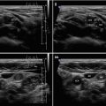
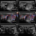
Fig. 10.2


(aa) A 51-year-old man with a large multinodular goiter. Medium-sized, complex, predominantly cystic nodule, size 38 × 26 × 19 mm and volume 10 mL in the RL and small, complex, predominantly solid nodule, size 20 × 19 × 15 mm and volume 3 mL in the LL. US overall view: complex, predominantly cystic nodule (arrowheads)—ovoid shape; inhomogeneous structure; hyperechoic; sporadic large cystic cavities (C); well-defined margin; complex, predominantly solid nodule (arrowheads)—round shape; coarse structure; hyperechoic; sporadic small cystic cavities (c); Tvol 34 mL, asymmetry—RL 21 mL and LL 13 mL; transverse, depth of penetration 4 cm. (bb) Detail of medium-sized complex nodule (arrowheads) in the RL: ovoid shape; inhomogeneous structure; hyperechoic; two large cystic cavities (C); small dotted and linear calcifications in the cavity wall (arrows); well-defined margin with halo sign; transverse. (cc) Detail of medium-sized complex nodule (arrowheads) in the RL: ovoid shape; inhomogeneous structure; hyperechoic; three cystic cavities (C), (c); small dotted and linear calcifications at the wall of cavity (arrows); well-defined margin with halo sign; longitudinal. (dd) Detail of small complex nodule (arrowheads) in the LL: round shape; coarse structure; hyperechoic; sporadic small cystic cavities (c); well-defined margin with thin halo sign; transverse. (ee) Detail of small complex nodule (arrowheads) in the LL: round shape; coarse structure; hyperechoic; sporadic small cystic cavities (c); well-defined margin with thin halo sign; longitudinal
Related posts:
Stay updated, free articles. Join our Telegram channel
Full access? Get Clinical Tree
