Localization: The axial skeleton is the preferred site. In the appendicular skeleton, the metaepiphyses are preferred. Very rare in bones of the hand and foot.
Clinical: Pain (usually relieved by rest), occasionally pathologic fracture (more often at a later stage). In advanced stages, there may be swelling, weight loss, anemia, bleeding diathesis, propensity to infections, hypercalcemic and hyperuricemic syndromes, subcutaneous myeloma masses, uremic renal insufficiency, jaundice, and ascites. Congestive heart failure, nephrotic syndrome, peripheral neuropathy, and carpal tunnel syndrome may result from amyloidosis. In 90 % of cases, serum and/or urinary immunoelectrophoresis shows a spike due to the excess of a monoclonal protein. Bence Jones proteinuria. Erythrocyte sedimentation rate is usually elevated. Elevated serum creatinine, hyperazotemia, renal clearances. Hypercalcemia (in advanced disease), due to massive osteolysis. Hyperuricemia is rather frequent, due to the release of purines and pyrimidines from dying neoplastic plasma cells. Anemia, leukopenia, and thrombocytopenia are due to substitution of bone marrow by the tumor.
Imaging: Radiographic skeletal changes remain absent, until the proliferative medullary disease has caused enough bone resorption.
(1) Diffused osteoporosis (osteopenia), particularly seen in the spine; (2) “punched-out” round lytic areas without associated osteoblastic changes, with sharp limits; no rim of sclerosis around the osteolyses; (3) large or massive osteolytic tumors (trabeculated, honeycombed, or even bubbly).
The cortices are thinned or canceled, and extension of the tumor in the soft tissues is frequent, particularly in the ribs and spine. Pathologic fractures are frequent. Periosteal reaction is absent. Rarely (1–2 %), multiple myeloma is osteosclerotic. This variant affects a slightly younger age and has fewer tumor cells, lower levels of monoclonal proteins, and more elevated serum alkaline phosphatase. It reflects a less aggressive infiltrative process, which permits time for osteoblastic reaction. MRI is the most sensitive to discover diffused and nodular disease in cancellous bone. Isotope bone scan is usually negative or scarcely positive.
Bone marrow aspiration. If necessary, it should be repeated from multiple sites. It is diagnostic in about 90 % of cases of multiple myeloma. If the smear contains over 3–5 % of plasma cells, diagnosis may be suspected. If it contains 10 %, the disease is very likely. If the plasma cell content is over 10–20 % and particularly if immature or anaplastic elements are seen, the diagnosis is certain.
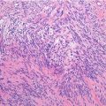
Histopathology: The myelomatous tissue is soft to gelatinous, grayish to dark red. Paramyloid substance may form grossly appreciable osseous or soft tissue masses, with a homogeneous and gelatinous appearance, yellowish gray or pink. It is composed by cells differentiating in plasma cells and producing a specific protein, but no matrix nor eliciting any fibrous or bone reactive tissue around them.
Histologically myelomatous tissue is composed by regular sheets of round to polygonal cells, separated from each other, without any matrix around them.
Differentiated myeloma: Plasma cells are mature, small, with abundant basophilic cytoplasm, a clear perinuclear halo (Golgi apparatus), and a round eccentric nucleus with chromatin clumping giving the “leopard skin” appearance. There is a moderate pleomorphism, some cells have two or more nuclei, and mitoses are rare.
Intermediate myeloma: Pleomorphism is more prominent; cells have a larger, frequently central hyperchromatic nucleus, often without the perinuclear halo. The nucleolus is more evident. Mitoses are not rare, both typical and atypical.
Anaplastic myeloma: Many cells have large, multiple, and bizarre nuclei; pleomorphism and hyperchromia are intense, nucleoli are prominent, and plasma cell features are not clearly evident.Related posts:

Stay updated, free articles. Join our Telegram channel

Full access? Get Clinical Tree