Musculoskeletal
QUESTIONS
1a Pictured here is a normal long (left) and short (right) axis image of the Achilles tendon. Which of the following best describes the ultrasound appearance of a normal Achilles tendon?
|

A. Hyperechoic with a fiber-like echotexture
B. Hyperechoic with a fascicular echotexture
C. Hypoechoic with a fiber-like echotexture
D. Hypoechoic with a fascicular echotexture
View Answer
1a Answer A. The normal appearance of a tendon is hyperechoic with a fiber-like or fibrillar echotexture, noted by the hyperechoic linear echoes within the tendon. The fibrillar echotexture within tendon represents the endotendineum, which is a combination of connective tissue, elastic fibers, nerve endings, blood vessels, and lymph vessels. This appearance is best demonstrated on long-axis images (LAX), which is the image on the left. On long-axis images, the proximal portion of the structure is by convention placed on the left side of the image. The short-axis image (SAX) is the image on the right and is acquired 90 degrees to the long-axis image, that is to say it is acquired transverse to the long-axis image. On each image, the Achilles tendon is superficial or near the top of the image.
Reference: Jacobson JA. Fundamentals of musculoskeletal ultrasound, 2nd ed. Philadelphia, PA: Elsevier, 2013:1.
1b Pictured here is a normal long (left) and short (right) axis image of the gastrocnemius and soleus muscle. What creates the hyperechoic striations within the muscle?
|

A. Fascia
B. Muscle bundles
C. Musculotendinous junction
D. Fibroadipose septa
View Answer
1b Answer D. The normal appearance of muscle is hypoechoic with hyperechoic fibroadipose tissue, known as perimysium, intervening between the muscle bundles. The image on the left is the long-axis image whereas the image on the right is the short-axis image. In both images, the gastrocnemius muscle is more superficial whereas the soleus is deeper. Fascia separates muscles instead of being within the muscle. The musculotendinous junction is not included in the provided images.
Reference: Jacobson JA. Fundamentals of musculoskeletal ultrasound, 2nd ed. Philadelphia, PA: Elsevier, 2013:1.
1c Pictured here is a normal long (left) and short (right) axis image of the median nerve. Which of the following best describes the ultrasound appearance of a normal nerve?
|

A. Hyperechoic with a fiber-like echotexture
B. Hyperechoic with a fascicular echotexture
C. Hypoechoic with a fiber-like echotexture
D. Hypoechoic with a fascicular echotexture
View Answer
1c Answer D. The normal appearance of a nerve is hypoechoic with fascicular echotexture. Separating the individual hypoechoic nerve fascicles is the hyperechoic epineurium, a type of connective tissue. This is best demonstrated by the image on the right, which is the short-axis image, where the nerve is seen superficially in the middle of the image with a honeycomb appearance of hypoechoic nerve fascicles separated by hyperechoic epineurium. The image on the left is the long-axis image, where the nerve is the more hypoechoic superficial structure with more hyperechoic tendons immediately deep to it.
Reference: Jacobson JA. Fundamentals of musculoskeletal ultrasound, 2nd ed. Philadelphia, PA: Elsevier, 2013:1.
1d Pictured here is a normal anterior talofibular ligament. Which of the following best describes the ultrasound appearance of a normal ligament?
|

A. Hyperechoic with a fiber-like echotexture
B. Hyperechoic with a striated appearance
C. Hypoechoic with a fiber-like appearance
D. Hypoechoic with a striated appearance
View Answer
1d Answer B. The normal appearance of a ligament is hyperechoic with a striated appearance. The ligament may appear less hyperechoic compared to a tendon because they are often adjacent to hyperechoic fat, which makes the ligament appear less echogenic in comparison. Ligaments contain striations within them, are more compact than tendons, and connect two osseous structures. The image is a long-axis image with the fibula on the left and the talus on the right side of the image.
Reference: Jacobson JA. Fundamentals of musculoskeletal ultrasound, 2nd ed. Philadelphia, PA: Elsevier, 2013:1.
2a Pictured here is the normal appearance of hyaline articular cartilage of the femoral condyle of the knee. Which of the following best describes the ultrasound appearance of hyaline cartilage?
|

A. Hypoechoic, homogeneous
B. Hypoechoic, heterogeneous
C. Hyperechoic, homogeneous
D. Hyperechoic, heterogeneous
View Answer
2a Answer A. The normal sonographic appearance of hyaline articular cartilage is homogeneously hypoechoic. As chondromalacia develops, the appearance may change, typically becoming heterogeneous.
Reference: Jacobson JA. Fundamentals of musculoskeletal ultrasound, 2nd ed. Philadelphia, PA: Elsevier, 2013:1.
2b Pictured here is the normal appearance of fibrocartilage within the knee meniscus. Which of the following best describes the ultrasound appearance of fibrocartilage?
|

A. Hypoechoic, homogeneous
B. Hypoechoic, heterogeneous
C. Hyperechoic, homogeneous
D. Hyperechoic, heterogeneous
View Answer
2b Answer C. The normal sonographic appearance of fibrocartilage is hyperechoic and homogeneous. Fibrocartilage includes the meniscus of the knee and the labrum of the shoulder and hip. Though these structures may be partially visualized with ultrasound, this is not typically considered the preferred method of evaluation for these structures. Tears of the fibrocartilage may be seen as a hypoechoic cleft within the otherwise hyperechoic structure.
Reference: Jacobson JA. Fundamentals of musculoskeletal ultrasound, 2nd ed. Philadelphia, PA: Elsevier, 2013:1.
3 Pictured here is a split screen image of the patellar tendon and deep infrapatellar bursa in short axis. The image on the left is the standard image whereas the image on the right suffers from anisotropy artifact. Anisotropy can result if the probe is as little as how many degrees off axis from perpendicular to the target?
|

A. 5 degrees
B. 15 degrees
C. 25 degrees
D. 35 degrees
View Answer
3 Answer A. Anisotropy is an important artifact to consider when imaging ligaments and tendons. To avoid anisotropy, the ultrasound beam should be perpendicular to the target. If the beam is as little as 5 degrees off perpendicular to the target, the image may suffer from anisotropy artifact. This artifact causes the tendon or ligament to appear hypoechoic, which can mimic pathology such as tendinosis. To confirm the finding is due to anisotropy, simply angle the probe so the beam is once again perpendicular to the target; if the hypoechogenicity does not persist, it was the result of artifact.
Reference: Jacobson JA. Fundamentals of musculoskeletal ultrasound, 2nd ed. Philadelphia, PA: Elsevier, 2013:1.
4 Pictured here is a split screen image of the patellar tendon and deep infrapatellar bursa in long axis. The image on the left is the standard image whereas the image on the right was obtained with spatial compound sonography. What is a benefit of this technique?
|

A. Improves evaluation of deep structures
B. Improves tissue plane definition
C. Improves artifact from motion blur
D. Improves conspicuity of foreign bodies
View Answer
4 Answer B. The provided images are long-axis images of the patellar tendon, with a portion of the patella on the left side of the images and tibial tuberosity on the right side of the images. Spatial compound sonography incorporates sound beams from multiple different angles to form the image. This improves tissue plane definition; however, it can have a smoothing effect and is more likely to produce motion blur because the frames are compounded to produce the image. It may also reduce the conspicuity of a foreign body because it can reduce the artifact associated with the foreign body, thereby making it more difficult to visualize. In this example, the interface between the surface of the tendon and adjacent fat is more clearly visualized.
Reference: Jacobson JA. Fundamentals of musculoskeletal ultrasound, 2nd ed. Philadelphia, PA: Elsevier, 2013:1.
5 Pictured here is a split screen image of the patellar tendon and deep infrapatellar bursa in long axis. The image on the left is the standard image whereas the image on the right was obtained with harmonics. What is a benefit of this technique?
|

A. Improves evaluation of deep structures
B. Improves tissue plane definition
C. Improves artifact from motion blur
D. Improves conspicuity of foreign bodies
View Answer
5 Answer A. Tissue harmonic imaging allows harmonic frequencies produced during ultrasound beam propagation to help produce the image. The benefits of this technique are improved visualization of deep structures and improved visibility of the surface of joints and tendons. It may also help visualize the borders of a mass or tendon tear. In this example, the deeper structures and the surfaces of the bones are more clearly defined.
Reference: Jacobson JA. Fundamentals of musculoskeletal ultrasound, 2nd ed. Philadelphia, PA: Elsevier, 2013:1.
6 Pictured here is an extended field of view image of a portion of the quadriceps tendon and the full length of the patellar tendon in long axis. Why does the patella produce a reverberation artifact?
|

A. Small radius of curvature and a smooth surface
B. Small radius of curvature and a rough surface
C. Large radius of curvature and a smooth surface
D. Large radius of curvature and a rough surface
View Answer
6 Answer C. The patella is creating reverberation artifact because it has a large radius of curvature and a smooth surface. This may be referred to as a dirty shadow. If an object has a small radius of curvature or a rough surface, it may produce a clean shadow.
Reference: Jacobson JA. Fundamentals of musculoskeletal ultrasound, 2nd ed. Philadelphia, PA: Elsevier, 2013:1.
7 Pictured here are two images of the patellar tendon both obtained in long axis. The images were obtained with two different probes; otherwise all of the parameters remained the same between the two images. Which of the following statements best describes the probes used to produce the images?
|

A. The image on the left is obtained with a 17-MHz linear transducer.
B. The image on the left is obtained with a 9-MHz curvilinear transducer.
C. The image on the right is obtained with a 17-MHz linear transducer.
D. The image on the right is obtained with a 9 MHz curvilinear transducer.
View Answer
7 Answer C. The image on the left was obtained with a 9-MHz linear probe whereas the image on the right was obtained with a 17-MHz linear probe. The superficial structures in the image on the right are more optimally visualized, particularly the echogenic fibrils within the tendon. For musculoskeletal ultrasound, typically linear probes with frequencies >10 MHz are preferred. Lower frequency probes may be used to visualize deeper structures. Curvilinear probes may be used for deeper structures as well, particularly the hip.
Reference: Jacobson JA. Fundamentals of musculoskeletal ultrasound, 2nd ed. Philadelphia, PA: Elsevier, 2013:1.
8 Pictured here is a split screen of two images of the patellar tendon both obtained in long axis. Which of the following techniques was used to acquire the images?
|

A. The image on the right is optimized for deeper structures with harmonics.
B. The image on the right is optimized for superficial structures with spatial compound sonography.
C. The image on the left is optimized for deeper structures with a 9 MHz probe.
D. The image on the left is optimized for superficial structures with proper placement of the focal zones.
View Answer
8 Answer D. The image on the left is optimized for superficial structures whereas the image on the right is optimized for deeper structures, both based on the position of the focal zones. The patellar tendon in the left image is optimally visualized, whereas the tendon in the image on the right is not. Neither image was obtained with harmonics. Both images were obtained with a 17-MHz probe. It is important to always adjust the focal zones over the target to be imaged.
Reference: Jacobson JA. Fundamentals of musculoskeletal ultrasound, 2nd ed. Philadelphia, PA: Elsevier, 2013:1.
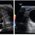
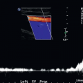
9 Pictured here is a split screen of two images of the common extensor tendon of the elbow. The image on the left is the standard image whereas the image on the right is with strain elastography. Which of the following statements best describes tendon elastography in this case?
|

A. The tendon is displaced more than the subcutaneous tissues.
B. The tendon is displaced less than the subcutaneous tissues.
C. The tendon is displaced the same as the subcutaneous tissues.
D. The tendon is displaced less than the bone.
View Answer
9 Answer B. Strain elastography measures the elastic properties of tissue. The degree of elasticity of a tissue is determined by the displacement of each structure within the box relative to one another when they are placed under strain from the ultrasound probe. The less a tissue is displaced, the harder or stiffer it is. The color scale can differ for each ultrasound manufacturer; in this case, the scale to right shows that SF (soft) is red whereas HD (hard) is blue. Structures that are hard are not displaced as much as soft structures. In this case, the tendon is hard relative to the subcutaneous tissues, which are soft. Therefore, the tendon is displaced less under strain than the subcutaneous tissues.
Reference: Jacobson JA. Fundamentals of musculoskeletal ultrasound, 2nd ed. Philadelphia, PA: Elsevier, 2013:1.
10 The two most common patient positioning techniques used to image the supraspinatus tendon are the Crass and modified Crass positions. Why are these techniques used?
A. Allow for tendon movement out from under the acromion
B. Allow for easiest probe placement
C. Allow for greatest patient comfort
D. Allow for greatest technologist control of the probe
View Answer
10 Answer A. The purpose of the Crass and modified Crass position is to bring the supraspinatus tendon out from under the acromion so it can be visualized. In the neutral position, the acromion creates shadowing, which obscures a large portion of the supraspinatus tendon leaving it incompletely evaluated. The Crass technique requires the patient to put the ipsilateral hand behind the back, which brings the greater tuberosity anterior. The modified Crass technique requires the patient to put the ipsilateral hand on the ipsilateral hip or as if it is in his or her back pocket. These techniques may create some discomfort for the patient, particularly in the setting of a tendon tear.
Reference: Jacobson JA. Fundamentals of musculoskeletal ultrasound, 2nd ed. Philadelphia, PA: Elsevier, 2013:8-15.
11 Pictured here are long-axis (left) and short-axis (right) images of a normal left rotator cuff tendon with labels. Which label is properly matched with the correct anatomic structure?
|

A. A—Trapezius muscle
B. B—Subscapularis tendon
C. C—Hyaline articular cartilage
D. D—Lesser tuberosity
View Answer
11 Answer C. The two images were obtained in the modified Crass position, with the ipsilateral hand positioned behind the patient’s back in the patient’s back pocket. This position brings the supraspinatus tendon out from under the acromion so it can be visualized; otherwise the acromion shadow would obscure its evaluation. One helpful way to understand the anatomy depicted in the images is to realize that the long-axis image is similar to the appearance of a coronal MRI image whereas the short-axis image is similar to a sagittal MRI image. The structures, which are typically seen from superficial to deep, include the skin, subcutaneous tissue, deltoid muscle, subacromial subdeltoid bursa, supraspinatus and/or infraspinatus tendon, hyaline articular cartilage, and humeral cortex. In this case, A represents the deltoid muscle, B the supraspinatus tendon, C the hyaline articular cartilage, and D the humeral cortex of the greater tuberosity. The hyaline articular cartilage is the hypoechoic structure overlying the humeral cortex, and its surface is noted by the echogenic reflection. The normal subacromial subdeltoid bursa either is not visible or is a thin hypoechoic structure between the deltoid muscle and the tendon. When the bursa contains fluid, such as in bursitis or a full-thickness rotator cuff tear, it will distend and become visible. The long-axis image will demonstrate either the supraspinatus or infraspinatus tendon whereas the short-axis image typically will demonstrate both tendons. The structure labeled E in the short-axis image is the long head of the biceps tendon. The subscapularis tendon, which inserts onto the lesser tuberosity, is visualized with a different technique whereas the teres minor tendon is not typically evaluated with a routine shoulder ultrasound.
Reference: Jacobson JA. Fundamentals of musculoskeletal ultrasound, 2nd ed. Philadelphia, PA: Elsevier, 2013:2-18.
12 Pictured here are the long-axis (left) and short-axis (right) images of the right supraspinatus tendon. What is the most likely diagnosis?
|

A. Normal tendon
B. Tendinosis
C. Partial-thickness tear
D. Full-thickness tear
View Answer
12 Answer C. The first thing to notice in the images is the focal hypoechogenicity of the tendon on both the long and short-axis images abutting the hyaline articular cartilage. This focal hypoechoic area persisted even with changing the angle of the transducer, suggesting it was not the result of anisotropy artifact. Tears of the rotator cuff tendons are divided into partial-thickness and full-thickness tears. Partial-thickness tears are then subdivided into articular-sided tears (meaning the tear extends to the hyaline articular cartilage but not the bursa), intrasubstance tears (meaning the tear is within the tendon and does not extend to either the hyaline articular cartilage or the bursa), or bursal-sided tears (meaning the tear extends to the subacromial subdeltoid bursa but not the hyaline articular cartilage). In this case, the tear extends to the hyaline articular surface consistent with an articular-sided partial-thickness tear.
Reference: Jacobson JA. Fundamentals of musculoskeletal ultrasound, 2nd ed. Philadelphia, PA: Elsevier, 2013:18-32.
13a Pictured here are the long-axis (left) and short-axis (right) images of the right supraspinatus tendon. What is the most likely diagnosis?
|

A. Normal tendon
B. Tendinosis
C. Partial-thickness tear
D. Full-thickness tear
View Answer


13a Answer D. The images depict a tear of the supraspinatus tendon that extends from the bursal surface to the articular surface, consistent with a full-thickness tear of the entire tendon. The tendon is retracted several centimeters. There is also a full-thickness retracted tear of the infraspinatus tendon.
Related posts:
Stay updated, free articles. Join our Telegram channel
Full access? Get Clinical Tree
