Musculoskeletal Infectious and Inflammatory Disorders


Musculoskeletal Infectious and Inflammatory Disorders


Clara L. Ortiz-Neira

Jennifer Stimec

Marcia Torre Moreira

Andrea S. Doria


INTRODUCTION

Timely and accurate diagnosis of musculoskeletal infectious and inflammatory disorders remains a challenge in infants and children particularly solely based on clinical grounds. In many cases, mimickers such as malignant tumors or benign but aggressive-appearing bone infarcts may constitute a diagnostic dilemma in the pediatric population. Musculoskeletal infections and inflammatory disorders can affect cartilage, synovium, muscles, bones, and joints.

Imaging evaluation that can visualize abnormalities in all of these anatomic structures is often necessary for reaching a definitive diagnosis in pediatric patients with various underlying congenital and acquired musculoskeletal infectious and inflammatory disorders. In this chapter, currently available imaging modalities and techniques, relevant clinical and laboratory manifestations, characteristic imaging findings, and up-to-date management for pediatric musculoskeletal infectious and inflammatory disorders encountered in clinical practice are discussed.


IMAGING TECHNIQUES


Radiography

Radiography is generally the first imaging modality to be considered when infectious or inflammatory musculoskeletal processes are clinically suspected. It can be often useful for excluding other musculoskeletal causes of patients’ symptoms such as fractures and tumors. For evaluation of soft tissue infection, radiography is not necessarily required but may provide valuable information such as demonstrating the presence of soft tissue gas or foreign body. For assessment of bone infection, the sensitivity of radiography ranges from 43% to 75% and its specificity ranges from 75% to 83%.1 As a result, initial radiographic results are often negative particularly in the early stages of infectious and inflammatory musculoskeletal disorders. For example, radiographic evidence of loss of bone mineralization is not evident on radiographs until the loss reaches 30% to 50%.2,3,4,5 However, radiographs may provide subtle information on early findings of infectious and inflammatory musculoskeletal disorders, which include soft tissue swelling, focal osteopenia, subperiosteal resorption, focal radiolucencies (Fig. 22.1), and periosteal reaction.2 Therefore, radiographs may provide clues on underlying pathologic conditions and guide subsequent imaging studies for further investigation. In addition, in cases of chronic infectious and inflammatory disorders, radiographs can be helpful in determining the presence, progression, healing, and complications of the process (Figs. 22.2, 22.3 and 22.4). In more advanced cases, new periosteal bone formation and cortical destruction can also be detected on radiographs.


Ultrasound

Because of its unique ability to clearly visualize soft tissues without potentially harmful radiation, ultrasound (US) is an attractive imaging modality for use in the pediatric population. US examinations are usually easily tolerated even by infants and young children with parents often holding them for maximal comfort. High-resolution musculoskeletal US requires the use of high-frequency linear transducers (10 to 15 MHz), which can provide optimal visualization of superficial and small structures in the pediatric population. If color Doppler is added to the technique, it can help evaluate hyperemia around the periosteum, synovium, and surrounding soft tissue abscesses often associated with infectious and inflammatory disorders.


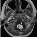
FIGURE 22.1 Radiography. Five-year-old girl who presented to the emergency room with knee pain and limping. A: Initial frontal radiograph shows a subtle lucency (arrow) in the lateral epiphysis of the distal femur. B: Follow-up MRI examination performed 10 days after onset of symptoms shows progression of the epiphyseal abnormality, which now abuts the physis and presents with further bony destruction. Increased signal consistent with bone marrow edema is seen on the coronal short tau inversion recovery (STIR) image (B), which corresponds to low signal on coronal T1-weighted MR image (C) in the epiphysis surrounding the focal bone abscess. Axial STIR (D) and axial post-contrast fat-suppressed T1-weighted (E) MR images show debris levels (arrow) within the intraosseous abscess, which does not enhance centrally. Peripheral rim enhancement is noted in the abscess (E).


FIGURE 22.2 Radiographic sequelae of osteomyelitis. Pathologic fracture through the proximal humerus in a preschool child with sickle cell disease and features of chronic osteomyelitis include diffuse cortical thickening of the diaphysis and bone expansion.

US can show soft tissue abnormalities as early as 2 days after the onset of symptoms.6 Although US is not typically used for evaluation of cortical bone abnormalities, periosteal elevation or subperiosteal collections presenting as a hypoechoic layer of purulent material can sometimes be evaluated with US (Fig. 22.5). US is useful for diagnosing and following up on soft tissue abscesses, for identifying precipitating factors such as foreign bodies or fistulae, for detecting effusion when transient or septic arthritis is clinically suspected (Fig. 22.6), and for guiding percutaneous drainage of fluid collections associated with infection for therapeutic purposes.4 In addition, US can be useful for evaluating regions that are complicated by orthopedic instrumentation and therefore might not be well seen with computed tomography (CT) or magnetic resonance imaging (MRI). Furthermore, US can be a valuable imaging modality in pediatric patients for whom MRI is contraindicated.


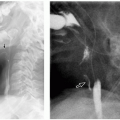
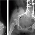
FIGURE 22.3 Radiographic sequelae of osteomyelitis. One-month-old infant boy with a focal lucency (arrow) in the left proximal femoral metaphysis that is subluxed superolaterally (A). Four years later, follow-up radiograph (B) demonstrates progression with hip dislocation, complete destruction of the epiphysis, and remodeling of the metaphysis.


Computed Tomography

Computed tomography (CT) is an excellent imaging modality for assessing cortical bone and for detecting gas in soft tissues. However, because it is radiation bearing, the use of CT is currently limited in infants and children with underlying musculoskeletal infectious or inflammatory disorders. Overall, MRI is a preferred imaging modality over CT for evaluation of bone or soft tissue infection or inflammation. However, CT may be the only option for infants and young children when sedation is contraindicated and thus, MRI cannot be performed.4


FIGURE 22.4 Radiographic sequelae of osteomyelitis. Chronic sequela of meningococcemia in infancy. Frontal radiograph of both knees obtained 3 years after the meningococcemia episode (A) shows multiple metaphyseal and epiphyseal lucencies with irregularity of the growth plates. Scanogram of lower extremities obtained a decade later (B) shows premature physeal fusion with resultant length discrepancy. Note the fibular bowing (arrow) due to tethering from the severely shortened tibia.

The currently available postprocessing techniques including multiplanar and 3D reconstructed CT images allow delineation of even the most subtle osseous changes such as early erosions due to infection or inflammation. CT can also clearly show abnormal thickening of the affected cortical bone, sclerotic changes, encroachment of the medullary cavity, and small draining sinuses in chronic osseous infections. In addition, CT can assist in guiding bone biopsies for diagnosis and draining deep abscesses for treatment. For evaluation of bones alone, intravenous (IV) contrast is not usually needed. However, the use of IV contrast can help better assess the presence and extent of associated soft tissue abnormalities such as abscess formation.


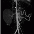
FIGURE 22.5 Ultrasound. Normal frontal radiograph of right tibia and fibula in a child with clinical suspicion for osteomyelitis (A). Ultrasound performed on the following day (B) shows a large subperiosteal abscess (arrow) along the proximal fibular metadiaphysis. Corresponding color Doppler ultrasound imaging (C) shows hyperemia in the adjacent soft tissues. The normal contralateral left side is included for comparison.


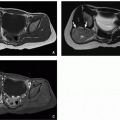
FIGURE 22.6 Ultrasound and nuclear medicine. Infant boy who presented with fever, irritability, and lack of mobility of the left lower extremity. Longitudinal grayscale ultrasound (A) shows large effusion (calipers) in the left hip. Ultrasound image of the contralateral asymptomatic hip (B) is shown for comparison. Bone scan performed on the same day of the ultrasound scan (C) demonstrates increased uptake in the proximal femur and left hip joint (arrow) suggesting septic arthritis. Aspiration of pus from the hip joint and culture of the material confirmed the diagnosis.


Magnetic Resonance Imaging

Magnetic resonance imaging (MRI) is currently the only imaging modality with the ability to simultaneously assess all relevant anatomic structures in the setting of musculoskeletal infectious or inflammatory disorders. It is the study of choice when a focal or multifocal area of osteomyelitis is suspected. For detecting soft tissue abnormalities, its specificity is up to 97%7 and its sensitivity, up to 100%.8 MRI evaluation can reliably differentiate acute from chronic infection.9 However, MRI may not be helpful in distinguishing infectious from noninfectious inflammatory processes. Recently, screening for multifocal noninfectious conditions with whole-body MRI has shown promising results.10 Whole-body MRI has a great potential to become evolved into an established and sensitive imaging modality for the diagnostic workup of pediatric patients with underlying multifocal infectious or inflammatory disorders. With the unique advantage of providing a comprehensive single evaluation of the entire body in a reasonable time without IV contrast or radiation, it is particularly beneficial in the pediatric population. Disadvantages of MRI include its occasional inability to differentiate infectious
from reactive inflammation as well as its difficulty with evaluating regions with metallic implants.

The basic MR imaging protocol for investigation of musculoskeletal infectious and inflammatory disorders includes T1 and T2/short tau inversion recovery (STIR) performed in orthogonal planes. T1-weighted MR imaging is useful for assessment of anatomy and bone marrow. A 3D image acquisition using T1 gradient sequence is useful in the detection of bone erosions, because it allows the generation of multiplanar reconstruction, which adds diagnostic confidence. Fluid-sensitive imaging sequences (T2/STIR) can delineate contrast between normal and abnormal tissue. A gradient echo sequence can be useful to identify hemosiderin, seen as blooming artifacts. The use of gadolinium helps delineate the extent of bone marrow abnormality, differentiate devitalized from vascularized bone, and demonstrate the extent of soft tissue abscesses (Fig. 22.7) and sinus tracts.11


Nuclear Medicine

Currently, because of the availability of MRI, nuclear medicine studies are rarely performed for evaluation of infectious or inflammatory disorders particularly in pediatric patients. Nevertheless, they may provide valuable information for children for whom CT or MRI is contraindicated.

Nuclear medicine studies can be of value for evaluating multifocal infections or as a screening imaging modality although they are limited mainly by low spatial resolution. Several nuclear medicine studies are currently available for evaluating pediatric musculoskeletal infectious and inflammatory disorders including technetium-99m methylene diphosphonate (99mTc) bone scan, gallium-67 citrate scan, and indium-111-labeled white blood cell (WBC) scan.

A 99mTc bone scan is usually performed when the area of infection is not well established clinically and the presence of a bone infection is uncertain. It shows elevated blood flow and abnormal increased deposition of tracer at the site of infection (Fig. 22.8). The tracer accumulates in areas of increased vascularity as well as in areas of increased physiologic activity in direct proportion to the destruction of the bone and osteoblastic healing activity. Triple phase (angiographic, bloodpool, delayed-phase) bone scan can be used when the site of acute osteomyelitis is not known or when multifocal points of infection are suspected.2 A diagnosis of acute osteomyelitis requires tracer activity in all three phases of bone scan.2 Bone scan is highly sensitive (sensitivity of 85% to 100%) but not particularly specific (specificity of 54% to 96%) for detecting osteomyelitis.1,12 Both sensitivity and specificity of bone scans can be increased with the use of pinhole collimators and single photon emission computed tomography (SPECT).13 Because of its variable specificity, bone scan may not be able to distinguish among infection, tumor, and trauma. Furthermore, there can be initial false-negative results secondary to ischemia from vascular compression and thrombosis as well as obscuration of subtle abnormal uptake at the metaphysis by normal high physeal uptake.11,14

Indium-111-labeled WBC scan is used in the diagnostic evaluation of occult infections when other imaging modalities are either contraindicated or noninformative. Labeled leukocytes migrate to and accumulate at sites of inflammation, which are then visualized on nuclear medicine imaging.15 111In-labeled WBC scans detect localized inflammation but do not clearly distinguish between infectious and noninfectious inflammatory processes.15 They have been used in the diagnostic evaluation of fever of unknown origin, prosthetic joint infections, and vascular graft infections.

Gallium-67 citrate scan is more specific than 99mTc bone scan for assessment of chronic osteomyelitis, but false positives occur in conditions such as healing fractures, noninfected prosthesis, and tumors.16 Gallium scan uses radiogallium, which attaches to transferrin and leaks from the bloodstream to the regions of inflammation showing increased isotope uptake in sterile inflammatory conditions, infection, and malignancy.16 It is often performed in conjunction with bone scan. One limitation of the use of gallium scan is that it does not show bone detail particularly well and may not distinguish well between bone and nearby soft tissue inflammation. In addition, gallium scan involves a higher dose of radiation and requires a longer scanning time than does 99mTc bone scan, making it less appropriate for use in the pediatric population. Gallium scan can take up to 48 hours to be completed, whereas bone scan typically takes up to 3 hours with a delayed view at 24 hours, if necessary.16


Fluorodeoxyglucose Positron Emission Tomography

Fluorodeoxyglucose positron emission tomography-CT (FDG PET-CT) is currently widely used for pediatric patients with cancer but is rarely used to diagnose bone infections. However, its use has been demonstrated in evaluating multifocal areas of infection in chronic osteomyelitis.10


BONE INFECTIONS


Bacterial Agents


Osteomyelitis

Osteomyelitis is a common pediatric infection that affects cortical bone and bone marrow. It has an incidence of 1 in 5,000 pediatric cases and causes considerable morbidity and sequelae in the pediatric population.17 Approximately 50% of bone infections occur in patients younger than 5 years.18,19,20


Clinical and Laboratory Findings

In the pediatric population, the symptoms of acute osteomyelitis generally include fever, local tenderness, and swelling. Symptoms in the neonate may be far more subtle, manifesting only as limited limb motion. Inflammatory markers such as C-reactive protein and erythrocyte sedimentation rate are elevated in 80% to 92% of cases.19 The WBC count is usually high in musculoskeletal infections but can be normal in 40% of osteomyelitis patients. Biopsy specimens may lack diagnostic findings, and they have variable success in recovering a responsible organism, which occurs in only 48% to 85% of specimens submitted for culture.19 The low specificity of the clinical and laboratory findings makes imaging a crucial tool in the diagnosis of acute osteomyelitis.


Etiopathogenesis

Hematogenous infections of osteomyelitis are primarily bacterial in origin, with Staphylococcus aureus (S. aureus) being the most common organism followed by Streptococcus species, pneumococcus species, Haemophilus influenzae, and less commonly methicillin-resistant S. aureus (MRSA), gram-negative (Kingella kingae [K. kingae]) bacteria, mycobacteria (tuberculosis), fungi, parasites, and viruses.22

In MRSA infections, affected pediatric patients heal slowly and are more prone to develop sequelae such as limb shortening, coxa plana, and coxa valga.22 Pseudomonas infection is more commonly seen in IV drug users and in patients with diabetes and is usually concurrent with other infections such as those caused by S. aureus, Streptococcus, Escherichia coli, and Klebsiella. Other underlying conditions predisposing to bone infections are meningococcemia, sickle cell disease, immunodeficiency, and varicella.19 In the past, infections by H. influenzae were common in children younger than 2 years of age.23 However, vaccination has significantly decreased the incidence of H. influenzae. Emphysematous osteomyelitis has been described in monomicrobial and polymicrobial bone infections and is characterized by the presence of gas in the bone marrow. Monomicrobial infections are usually hematogenous, whereas polymicrobial infections are usually related to the contiguous spread from infected soft tissues.20


FIGURE 22.10 Modes of spread of infection. A: Infant blood supply demonstrates continuity of the metaphyseal and epiphyseal vasculature through the presence of transphyseal vessels, allowing a pathway for spread of infection. B: In the preschool child and adolescent, the transphyseal vessels are no longer patent; thus, the physis acts as a barrier to the spread of infection into the epiphysis.


Acute Osteomyelitis

Acute hematogenous osteomyelitis is the most common form of bone infection in the pediatric population. Acute osseous infection results in extensive inflammatory response and leads to increased intraosseous pressure, blood stasis, thrombosis, and subsequent bone necrosis.5,21

In early stages of osteomyelitis, ill-defined bony radiolucencies are often observed on radiographs. Later on, cortical destruction, periosteal elevation, and spread of infection into the adjacent soft tissues are typical. Periosteal elevation accompanying osteomyelitis is more pronounced in children than in adults because of the loose attachment of the periosteum to bone in children.5

On MRI, acute osteomyelitis is represented by areas of bone marrow edema, most frequently seen in the metaphysis of the long bones (Table 22.1). Bone marrow edema, which
can be an initial sign of osteomyelitis, appears hypointense on T1-weighted MR images and hyperintense on T2-weighted MR images. With the administration of contrast, infected bone marrow typically enhances.16 MRI may also demonstrate regional periosteal reaction, which is caused by disrupted vascular connections that elevate the periosteum and cause new layers of periosteum (involucrum) to form.24 Spread of infection with involvement of soft tissues is manifested as increased signal intensity on T2-weighted MR images. When an abscess forms within the bone, the marrow signal intensity becomes heterogeneous, and with contrast administration, the rim of the abscess can enhance (Fig. 22.7).


TABLE 22.1 Osteomyelitis: Definitions


Disorder


Definition


Acute osteomyelitis


Acute infection of bone and bone marrow; usually caused by bacteria from hematogenous infection.


Subacute osteomyelitis


Bone infection lasting >4 weeks and <3 months; classic presentation, a Brodie abscess.


Chronic osteomyelitis


Bone infection lasting at least 3 months; sequestrum, involucrum, and cloaca can be present in this phase of the infection.


Brodie abscess


Subacute osteomyelitis; well-defined lytic lesion that corresponds to bone necrosis surrounded by a fibrous granulation tissue.


Sequestrum


A piece of nonresorbed devascularized infarcted bone surrounded by new bone.


Involucrum


Periosteal new bone formation caused by chronic infection or inflammation over necrotic bone.


Cloaca


An opening tract from the cortex of bone.


Chronic recurrent multifocal osteomyelitis (CRMO)


A noninfectious, inflammatory disorder of bone.


Subacute Osteomyelitis

Subacute osteomyelitis is considered when the diagnosis of infection is made between 1 and 4 weeks from the onset of symptoms.19 It remains localized either by the low virulence of the organism or increased host resistance.21 The development of subacute osteomyelitis seems to depend on the interplay between the infecting bacteria and the immune system of the host and represents a favorable host-pathogenic response.9

If the infection continuously evolves, it can extend into the bone marrow, generating an intraosseous abscess called Brodie abscess (Table 22.1; Fig. 22.11). Typical radiographic features of Brodie abscess, which is usually located in the metaphysis, consist of a localized destructive lucent bone lesion with a surrounding sclerotic rim with variable thickness. On MRI, Brodie abscess has a characteristic target appearance with four distinct layers. A central abscess cavity appears hypointense on T1-weighted and hyperintense on
T2-weighted MR images. Surrounding the cavity is a layer of highly vascularized granulation tissue that appears isointense on T1-weighted and hyperintense on T2-weighted MR images (Fig. 22.12). This layer is best visualized on contrast-enhanced MR images because the halo enhancement corresponds to granulation tissue. Next is a fibrous layer that demonstrates low signal intensity on all MR sequences. The outer halo is composed of a peripheral rim of endosteal reaction and sclerosis that has low signal on T1-weighted and T2-weighted MR images.25 The halo enhancement on postcontrast MR images corresponds to granulation tissue. The bone marrow edema that usually surrounds the abscess
appears hypointense on T1-weighted and hyperintense on T2-weighted MR images.10,26 Brodie abscess is managed with curettage and often lengthy antibiotic treatment.27


FIGURE 22.11 Brodie abscess. Frontal radiograph (A) of the tibia and fibula of an 8-year-old boy with a Brodie abscess shows a lytic tibial diaphyseal lesion (arrow) with associated periosteal reaction best seen on the axial CT image (B). (Continued)


FIGURE 22.11 (Continued) Corresponding histologic slides show pus surrounding resorbing scalloped necrotic bone (C, hematoxylin and eosin, original magnification, 600×), and clusters of gram-positive cocci (arrows) seen within the purulent material (D, Brown-Brenn stain, original magnification, 600×). This infection was subacute, having been partially treated by the parents with leftover antibiotics from their medicine cabinet before seeking medical attention.


FIGURE 22.12 Brodie abscess. Frontal radiograph of the left femur (A) shows a geographic lytic lesion (arrow) with sclerotic borders in the distal metaphysis. Sagittal proton density-weighted MR image of the corresponding knee (B) demonstrates the typical target appearance of a Brodie abscess (asterisk) with a hypointnse center (intraosseous abscess), a hyperintense inner ring of vascular granulation tissue (arrow) and an outer ring of low signal fibrotic tissue and sclerosis.


FIGURE 22.13 Features of chronic osteomyelitis. Frontal radiograph of the tibia and fibula (A) shows diffuse sclerosis, remodeling, and periosteal reaction extensively involving mainly the left tibia (arrows) of an 8-month-old infant boy representing chronic osteomyelitis. Frontal radiograph of the region of interest obtained 4 years later (B) demonstrates multifocal bone bridging resulting in overall shortening of the left tibia (arrow), marked length discrepancy, and varus alignment, as complications.


Chronic Osteomyelitis

Chronic osteomyelitis can result from an untreated acute infection or a low-grade continuous infection persistent for at least 3 months, and most frequently, lasting 6 months or longer.28 It occurs more commonly in developing countries and is associated with significant morbidity and sequelae (Table 22.1).29 Because of its insidious onset, mild symptoms, lack of systemic reaction, and often inconsistent supportive laboratory data, chronic osteomyelitis may mimic various benign and malignant conditions, resulting in delayed diagnosis and treatment.30 Sequelae of chronic osteomyelitis include physeal destruction with growth arrest, angular deformities, ankylosis, and leg-length discrepancies (Fig. 22.13).31

Imaging features of chronic osteomyelitis include bone sclerosis (Fig. 22.13) with underlying bone resorption and cystic changes.21 Alternatively, complications such as the formation of a sequestrum (necrotic bone with new bone applied to its surface), involucrum (thick periosteal bone formation surrounding a sequestrum), or cloaca (draining fistula between bone marrow and periosteum) (Fig. 22.14) may occur. At this chronic stage, MRI findings of osteomyelitis include heterogeneity of the bone marrow signal on
T1- and T2-weighted MR images, with areas of increased and decreased signal intensity. Gradient-echo MR sequences can be helpful for delineation of sequestra, periosteal reaction, and involucrum because susceptibility artifacts from mineralization are exaggerated. Areas of chronic fibrosis or dead bone (including sequestrum) have low signal intensity on both T1- and T2-weighted MR images, which do not enhance with contrast. A sequestrum may be surrounded by tissue that has high-signal intensity on T1- and T2-weighted MR image and enhances with contrast (Fig. 22.14). An involucrum appears as cortical thickening with healing (Table 22.1). If the infection persists, a sinus tract or cloaca can drain pus into the adjacent soft tissues (Fig. 22.15).


FIGURE 22.14 Features of chronic osteomyelitis. Frontal radiograph of the distal humerus of a 12-year-old boy who had undergone pinning of a supracondylar fracture 2 years previously (A) shows sclerosis, cortical thickening, and cloaca (arrow). Coronal fat-suppressed T2-weighted MR image (B) demonstrates an intraosseous abscess with a cloaca (arrow) extending superolaterally into a soft tissue abscess. (Continued)


FIGURE 22.14 (Continued) A low-signal sequestrum (arrow) is seen centrally in the hyperintense osseous abscess on the axial fat-suppressed T2-weighted MR image (C). Axial T1-weighted MR image (D) depicts the cloaca (arrow) along the anterior cortex. Axial post-contrast fat-suppressed T1-weighted MR image (E) shows the path of the soft tissue component of the abscess (arrowheads) tracking through the anterior cortex superficially (arrow) dissecting into the subcutaneous soft tissues.

Pediatric patients with chronic osteomyelitis usually require further evaluation with bone scan and MRI in order to assess their response to treatment and chronic complications such as soft tissue abscess or fistula formation. In order to detect sequestra or cortical thickening in chronic stages of osteomyelitis for guiding biopsy or debridement, radiologists should consider using CT over MRI.16

In summary, MR imaging features of chronic osteomyelitis usually include the following:


  • Bone marrow edema with or without associated enhancement.


  • Focal cortical destruction and associated periosteal elevation.


  • Associated intraosseous or juxtacortical soft tissue abscesses or edema.


  • Cloaca (bone marrow to periosteum) or sinus tract formation.


  • Sequestrum or involucrum formation.


Oct 13, 2018 | Posted by in PEDIATRIC IMAGING | Comments Off on Musculoskeletal Infectious and Inflammatory Disorders

Full access? Get Clinical Tree

Get Clinical Tree app for offline access