Musculoskeletal Tumors

25
Musculoskeletal Tumors

Eugene C. Lin and Abass Alavi


Image Distinguishing Benign from Malignant Musculoskeletal Tumors


In a known bone or soft tissue lesion, positron emission tomography (PET) is of some value in determining whether the lesion is benign or malignant and in grading malignant lesions. In addition, bone or soft tissue lesions are detected incidentally on PET performed for other indications. The degree of fluorodeoxyglucose (FDG) uptake in the lesion can aid in differential diagnosis when correlated with conventional imaging modalities.


Benign versus Malignant Bone Tumors


Clinical Indication: C


  1. Low uptake. A low level of uptake suggests that a bone lesion is likely benign (although there are false-negative results in plasma-cytoma and low-grade chondrosarcoma).
  2. High uptake. A high level of uptake is less specific. Although lesions with high uptake are more likely to represent malignancy (primary or metastatic), high uptake can be seen in a large number of benign lesions (see Pitfalls section).
  3. Chondrosarcoma versus enchondroma1


    1. Chondrosarcomas (Fig. 25.1) usually have less uptake than other sarcomas but more uptake than enchondromas.
    2. PET cannot distinguish between benign tumors and grade I chondrosarcomas.
    3. Grade II and III chondrosarcomas have higher glucose metabolism than low-grade cartilage tumors.


      • A standardized uptake value (SUV) cutoff of 2.3 is helpful in differentiating grade II and III chondrosarcomas from low-grade tumors.

  4. Chondrosarcoma versus osteochondroma. Limited data suggest an SUV cutoff of 2.0 may differentiate benign from malignant osteochondromas.2

Accuracy


  1. PET. Sensitivity 93%, specificity 67%3


    • These results were obtained using tumor-to-background ratio of 3.0 as a positive result.

Pearls


  1. In general, malignant bone lesions have higher FDG uptake than benign lesions.
  2. Metastases have the highest uptake, usually more than primary malignant bone lesions.4


  3. However, benign bone tumors can have substantial FDG accumulation (> 2.0 SUV).


Pitfalls


  1. False-negatives.3,5 Low-grade chondrosarcoma, plasmacytoma, myxoid tumors
  2. False-positives.6,7 Giant cell tumor, chon-droblastoma, fibrous dysplasia, sarcoidosis, Langer hans cell histiocytosis, nonossifying fibroma, osteoblastoma, aneurysmal bone cyst, Paget disease (active), enchondroma, chondromyxoid fibroma, desmoplastic fibroma, brown tumor, fibro-osseous defects, osteomyelitis, bone infarct, acute or sub-acute fracture

Benign versus Malignant Soft Tissue Tumors


Clinical Indication: C

  1. Low uptake. Low FDG uptake is of limited value in differentiating benign from malignant soft tissue tumors, as it could be represent either a nonmalignant lesion or a low-grade sarcoma.
  2. High uptake. High uptake is more useful, as it usually indicates intermediate or high-grade malignancy. Although some benign lesions can have high uptake, radiographic correlation can often differentiate these lesions from sarcomas.


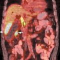
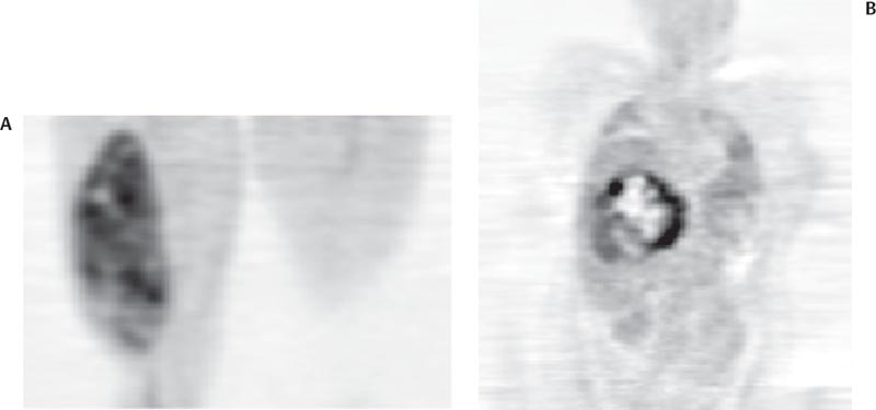
  3. Liposarcoma versus lipoma. Limited data suggest that an SUV cutoff of 0.81 may differentiate liposarcomas (Fig. 25.3) from lipomas.8


    image

    Fig. 25.3 Liposarcomas. Coronal positron emission tomography (PET) scans demonstrate fluorodeoxyglucose uptake in liposarcomas in the (A) thigh and (B) abdomen. The abdominal liposarcoma is heterogeneous with a standardized uptake value (SUV) of 11.7, consistent with a poor prognosis tumor. Given the heterogeneity of uptake, the PET scan would be helpful in guiding biopsy. The thigh liposarcoma has a much lower SUV of 2.7, consistent with a better prognosis tumor. However, there is some degree of heterogeneity, suggesting myxoid degeneration and a poorer prognosis than might be predicted by the SUV alone. (Courtesy of Janet Eary, MD, Seattle, WA.)


Accuracy


  1. PET. Sensitivity 92%, specificity 73%9


    • These results were obtained using qualitative interpretation.

Pearls and Pitfalls


  1. Tumor grade. FDG uptake correlates with tumor grade.10


    1. Lesions with SUV ≥ 1.6 are usually high grade.
    2. Lesions with SUV < 1.6 are usually low grade or benign.

  2. Benign lesions. Benign soft tissue lesions usually do not have substantial FDG uptake.11


    1. Lipomas and hemangiomas have the lowest uptake.
    2. False-positives. High uptake can be seen in giant cell tumor of the tendon sheath, sarcoid, desmoid, and schwannomas. Uptake in a hibernoma can mimic a liposarcoma.12

  3. Delayed imaging. Delayed imaging can help differentiate benign from malignant tumors, as malignant lesions show an increase in uptake on delayed images.


Image Evaluation of Known Musculoskeletal Tumors


PET is valuable in both osseous and soft tissue malignancies. The primary uses are staging, guiding biopsy, detecting recurrence, therapy response, and tumor grading.


Osteosarcoma and Soft Tissue Sarcomas14,15


Clinical Indication: B
The primary applications of PET are in guiding biopsy, therapy monitoring, and diagnosing local recurrence.


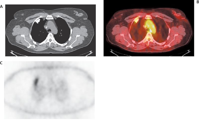
  1. Staging. PET is of limited value in staging osteosarcoma (Fig. 25.4).


    1. PET is less sensitive than bone scan for the detection of bone metastases from osteosarcoma.16


      image

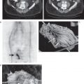
      Fig. 25.4 Osteosarcoma lung metastasis. (A) Computed tomography (CT) scan in a patient with osteosarcoma demonstrates a calcified right upper lobe lesion. (B) Axial positron emission tomography (PET)/CT scan demonstrates uptake in this lesion consistent with metastasis. (C) In this case, it is important to review the nonattenuation-corrected image to determine that the uptake is not artifactual secondary to the increased density of the lesion. Although PET can detect lung metastases from osteosarcoma, CT is more sensitive for this purpose.

      Only gold members can continue reading. Log In or Register to continue

      Stay updated, free articles. Join our Telegram channel

Sep 3, 2016 | Posted by in GENERAL RADIOLOGY | Comments Off on Musculoskeletal Tumors

Full access? Get Clinical Tree

Get Clinical Tree app for offline access