Myocardial Perfusion Imaging with PET, PET/CT, PET/MRI: Technical Advances and Future Applications
Myocardial Perfusion Imaging with PET, PET/CT, PET/MRI: Technical Advances and Future Applications
Vikas Veeranna, MD
Sharmila Dorbala, MD, MPH
▪ Introduction
Radionuclide myocardial perfusion imaging (MPI) remains the mainstay for the diagnosis, risk assessment, and management of patients with known or suspected coronary artery disease (CAD).1 MPI with radionuclide techniques can be accomplished with either single photon emission computed tomography (SPECT) or positron emission tomography (PET). With the improved image quality from higher spatial and temporal resolution, increased availability of PET scanners and radiotracers, and the ability to assess myocardial blood flow (MBF), PET MPI makes for an attractive alternative to SPECT MPI.2,3,4,5 and 6
PET is a noninvasive imaging modality that can be used to quantitatively assess minute biochemical pathways using radiotracers containing naturally occurring elements such as carbon, nitrogen, oxygen, and fluorine. Since its first use more than 50 years ago, PET has considerably evolved in terms of hardware, software, radiotracers, and clinical applications. Apart from relative and absolute quantitation of MBF, PET allows for the evaluation of myocardial metabolism and identification of ruptured or high-risk atherosclerotic plaques.2,3,7,8 and 9 Further, cardiac PET has been used for imaging inflammation, sympathetic innervation, and infiltrative diseases of the heart.3,10,11 and 12 With these technologic and clinical developments, PET may soon become the diagnostic modality of choice for the assessment of several cardiovascular diseases.2,3,5,10,13,14 and 15 Furthermore, hybrid imaging using separate scanners or integrated hybrid scanners provides an opportunity for advanced imaging combining anatomical, physiologic, and functional information. In this chapter, we will focus on the recent advances in radiotracers, technology, and some of the novel clinical applications for PET MPI.
▪ Advances in PET MPI
Myocardial Perfusion Radiotracers
Rubidium 82 or N-13 ammonia is used for clinical applications of PET MPI, while O-15 water is used for research applications.2,9,16 Rubidium 82 is generator produced increasing its availability for sites without a cyclotron, but is expensive and in limited supply. N-13 ammonia, due to its 9.96-minute half-life, requires a cyclotron in close proximity to the imaging center. Conventional cyclotrons require a large space (which may be limited in medical centers with space constraints) and capital investment. Thus, PET radiotracer availability is a major limitation for more widespread use of PET MPI and had led to the development of novel cyclotrons and novel radiotracers. The development of novel compact cyclotrons solely for the production of N-13 ammonia circumvents some of these issues. A compact (room size 150 square feet), point-ofcare, 12-MeV, self-shielded superconducting cyclotron, ION-12sc, was developed by Ionetix Corporation.17 Also, table-top cyclotron was developed by the University of Michigan,18 and a laser plasma accelerator developed by Berkeley National Laboratory19 will make N-13 ammonia more accessible for medical imaging.
Also, unlike O-15 water, rubidium 82 and N-13 ammonia are not completely extracted during first-pass circulation throughout the heart and not linearly taken up by the myocardium in relation to blood flow particularly during hyperemia. Further, the exercise stress is challenging with short-acting radiotracers such as rubidium 82 and N-13 ammonia PET MPI, limiting greater clinical applicability. These limitations have led to an interest in the development of perfusion tracers with superior extraction characteristics and tagged with F-18 for a longer half-life (110 minutes). These fluorinated radiotracers could be used for exercise PET perfusion imaging and shipped to various sites as unit doses, allowing for greater accessibility to a PET perfusion tracer. Several fluorinated PET perfusion tracers are under evaluation: (a) F-18-BMS-747158-02 (2-tert-butyl-4-chloro-5-[4-(2-(18F)fluoroethoxymethyl)-benzyloxy]-2H-pyridazin-3-one); (b) 2-tert-butyl-4-chloro-5-{6-[2-(2-18F-fluoroethoxy)-ethoxy]-pyridin-3-ylmethoxy}-2H-pyridazin-3-one(18F-BCPP-EF); and (c) 2-tert-butyl-4-chloro-5-[6-(4-18F-fluorobutoxy)-pyridin-3-ylmethoxy]-2H-pyridazin-3-one (18F-BCPP-BF).20 Of these, F-18 BMS compound now known as F-18 flurpiridaz has been most extensively evaluated.
Fluorine 18 flurpiridaz (F-18 flurpiridaz) is a novel cyclotronproduced radiotracer with a long half-life of 110 minutes. Although, produced by a cyclotron, due to its long half-life, it can be produced at regional cyclotrons and delivered to imaging centers as unit doses (similar to F-18 FDG). It binds to the mitochondrial complex I of the electron transport chain21 and is taken up by the heart due to high mitochondrial densities in the myocardium.2,22 Phase 1 clinical trials established safety and biodistribution of F-18 flurpiridaz in humans.23 This tracer has a short positron range, high first-pass extraction (>90% even at high flow rates), slow wash-out, and a low background uptake.22 These properties provide for a higher spatial resolution and make F-18 flurpiridaz an excellent tracer for flow quantitation. Indeed, in pig models, when compared to N-13 ammonia, F-18 flurpiridaz showed higher target-to-background activity ratios between the myocardium and the blood pool, lungs, and liver both at stress and rest accounting for the higher-quality images.22 Further, regional MBF and defect extent correlated closely with radioactive microspheres.22 F-18 flurpiridaz is also well suited for use with exercise stress testing due to its longer half-life.16
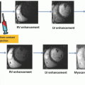
From Bengel FM, Higuchi T, Javadi MS, et al. Cardiac positron emission tomography. J Am Coll Cardiol 2009;54:1-15; Orbay H, Hong H, Zhang Y, et al. Positron emission tomography imaging of atherosclerosis. Theranostics 2013;3:894-902; Di Carli MF, Murthy VL. Cardiac PET/CT for the evaluation of known or suspected coronary artery disease. Radiographics 2011;31:1239-1254.
Results from a phase 2 trial showed F-18 flurpiridaz to be safe with superior image quality, improved diagnostic certainty, and more sensitivity compared to technetium (Tc)-99m SPECT MPI.24 This multicenter study included 143 patients who underwent Tc-99m SPECT MPI as well as F-18 flurpiridaz PET MPI and invasive coronary angiography (N = 86).24 For the detection of obstructive CAD by coronary angiography, when compared to SPECT MPI, PET MPI showed a significantly higher sensitivity (78.8% vs. 61.5%, p < 0.05) with no significant difference in specificity (76.5% vs. 73.5%, p = nonsignificant).24 Furthermore, in patients with angiographic CAD, when compared to SPECT MPI, PET MPI demonstrated a greater magnitude of reversible perfusion defects (90.8% vs. 70.9%, p < 0.01). These improved characteristics may be attributable to the physical characteristics of F-18-flurpiridaz including a higher extraction even at higher flow rates compared to traditional SPECT tracers.24
Compared to the available PET perfusion tracers, F-18 flurpiridaz has high target-to-background ratio, higher myocardial extraction and uptake, accurate MBF quantitation, low positron range, and longer half-life, allowing the tracer to be delivered in unit doses from regional cyclotrons. These features make this tracer an almost ideal PET tracer. However, the preliminary results of a phase 3 trial have shown similar high sensitivity but potentially lower specificity for the detection of CAD compared to SPECT MPI.25 More largescale clinical trials will be performed before it is FDA approved for clinical use (Table 1).
Technical Advances of PET, PET/CT, and PET/MR Systems
Several key technologic advances in software, hardware, and newer hybrid PET/CT and PET/MR systems have contributed to significant improvements in the performance characteristics of the present-day PET scanners.3,5Figures 22-1, 22-2 to 22-3 illustrate some of the important technical advances in PET imaging.
Software Advances
Software advances in PET MPI include iterative reconstruction algorithms, high-definition PET, and cardiac “motion freeze” imaging. Conventional PET systems used filtered back projection algorithms for image reconstruction along with corrections for randoms, scatter, dead time, attenuation, and decay. The drawback of filtered back projection is streak artifacts, which affect the visual interpretation, especially in patients with large body size.3 Iterative reconstruction algorithms improve image noise. These algorithms weigh the data based on their statistical quality and model the geometry of the imaging system such as intercrystal scatter and depth of interaction effects and nonuniform sensitivity along a line of response. These algorithms nearly eliminate streak artifacts and greatly improve the visual appearance of the image. However, these algorithms need high computational power to perform sufficient iterations particularly to avoid artifacts in regions with low radiotracer uptake.3 With the increased computer power of the current generation PET systems, iterative reconstruction is now the most commonly employed reconstruction protocol. Another improvement in image reconstruction is the introduction of high-definition PET with spatially variant 3D-specific point spread function (PSF), which can significantly improve spatial resolution of the images and signal-to-noise ratio providing high-quality images.26,27 Cardiac motion freeze (CMF) technique, which addresses loss of image resolution from cardiac motion on the static images, is another recent advancement. Initially developed for SPECT imaging, it can be applied to PET as well. CMF processing involves the tracking of left ventricular endo- and epicardial motion on the gated images and projecting the counts from all the cardiac phases to one single phase, usually end diastole. This resultant image has higher count statistics with spatial resolution similar to that of the end-diastolic image, and when applied with 3D-PSF, reconstruction algorithm provides a high-quality image free of blurring due to cardiac motion.28
Figure 22-1. Advances in PET technology. A: Basic positron emission tomography (PET) principle: a positron (e+) is emitted from the atomic nucleus together with a neutrino. The positron moves randomly through the surrounding matter, where it hits electrons (e-) until it finally loses enough energy to interact with a single electron. This process, called “annihilation,” results in two diametrically emitted photons with energy of 511 keV each. These photons are detected as coincidences in the detector ring of the PET camera. B: Traditional two-dimensional imaging (left) uses only coincidences that occur within the same axial detector ring. Adjacent detector rings are separated by septa. Advanced three-dimensional imaging (right) uses coincidences from all possible detector pairs. This increases sensitivity and count density but is demanding and requires correction for the higher amount of scatter and inhomogeneity at the axial edge of the field of view. (Reproduced with permission from Bengel FM, Higuchi T, Javadi MS, et al. Cardiac positron emission tomography. J Am Coll Cardiol 2009;54:1-15.)
Figure 22-2. Schematic showing the potential added value of time-of-flight (TOF) PET for localization of the event. With conventional (non-time-of-flight) imaging, the precise position of the emission event between the two opposing detectors is not known. All the pixels along the line of response must by incremented during reconstruction (right top). With TOF PET, the precise location of the event is better identified and the image can be accordingly reconstructed (right bottom). (Reproduced with permission from Lecomte R. Novel detector technology for clinical PET. Eur J Nucl Med Mol Imaging 2009;36(suppl 1):S69-S85.)
Figure 22-3. Multidimensional list-mode PET acquisition. Scanner coincidences are continuously recorded along with information about the time after the start of acquisition, the electrocardiographic signal, and the signal about breathing position (optional). Data can then be resampled in multiple formats at any time of the acquisition. A: High-count static images are reconstructed by summing all information after a predefined prescan delay (delay time after tracer injection). B: Dynamic imaging sequences are obtained by serial temporal sampling at different times after injection. This is used for tracer kinetic analysis. C: Electrocardiographically gated images are obtained at multiple phases of the cardiac cycle to assess ventricular function. D: Respiratory gated images can be obtained at different phases of the breathing cycle in order to correct for respiratory motion. (ED, end diastole; ES, end systole; EXSP, expiratory phase; INSP, inspiratory phase; PET, positron emission tomography). (Reproduced with permission. from Bengel FM, Higuchi T, Javadi MS, et al. Cardiac positron emission tomography. J Am Coll Cardiol 2009;54:1-15.)
Scintillation Crystals
At present, several different crystals—bismuth germanate (BGO); the newer gadolinium oxyorthosilicate (GSO), lutetium oxyorthosilicate (LSO), and lutetium yttrium orthosilicate (LYSO); and others—are used commonly in PET imaging.29 Although BGO has high stopping power and provides good detector efficiency at 511 keV, the slow decay time and low light output of BGO leads to relatively poor timing and energy resolution.3,5,30,31 and 32 Despite the lower stopping power, the main advantage of the newer crystals (GSO, LSO, and LYSO) is their significantly reduced dead time enabling 3D dynamic imaging for MBF quantitation. By their higher light output compared to BGO, these new scintillators also permit more crystal elements to be decoded per photomultiplier tube. These features have in turn contributed to development of higher quality images with PET MPI.3,5,30,32
Imaging Modes
Conventional PET scanners allow for imaging in a static mode, in an ECG-gated mode, or in a dynamic mode (Figure 22-3). The dynamic images are multiframe time sequence high temporal resolution images that enable assessment of radiotracer transit through the various cardiac chambers. Using a dynamic imaging sequence and tracer kinetic modeling techniques, absolute MBF, and physiologic or biochemical function can be estimated.3,33 Using conventional scanners, if MBF assessment is desired, the protocols are tailored such that the dynamic images are obtained over the first few minutes of the scan and a separate gated or static image acquisition is started. A recent advance in PET scanners is greater computer memory allowing for the option of list mode acquisition. List mode acquisition is a multiframe acquisition in relation to time and ECG. The advantage of list mode data acquisition is ability to reconstruct the data acquired during a single image acquisition into static images for perfusion assessment, gated images for function assessment, and dynamic images for MBF assessment.3,33 The ability to acquire images in a list mode has significantly enhanced PET MPI and has allowed for routine quantitation of MBF in all patients.
Semiconductor Detectors and Silicon Photomultiplier Tubes
Pixelated semiconductor detectors have been a recent advance in SPECT. Semiconductors directly convert the electronic signal into an image (allowing a compact system) and have a high sensitivity allowing for low radiation dose imaging. Their use is expanding to PET, and some of the next-generation PET scanners will incorporate semiconductor detector material cadmium telluride (CdTe). These detectors are compact and can be tightly packed and coupled one to one with the PET scintillation crystals.34 These semiconductor detectors appear to have slightly better spatial resolution and significantly better energy resolution and lower scatter compared to conventional PET.35,36 Also, conventional photomultiplier tubes are extremely sensitive to magnetic fields and limit the MR signal. For this reason, novel silicon-based solid-state sensors called avalanche photodiodes, which are insensitive to magnetic fields, have been developed for hybrid PET/MR systems. The silicon-based photomultiplier tubes also offer advantages of improved signal to noise and timing resolution allowing for time-of-flight (TOF) imaging.37
Time-of-Flight Imaging
TOF is the time difference between the two annihilation photons reaching their respective detectors 180 degrees apart.3,32 The coincidence electronics in the new advanced PET scanners with TOF electronics are capable of measuring the exact time interval between the two annihilation photons reaching the opposing detectors. The exact location of the annihilation is estimated by multiplying the difference in time with the speed of light along the coincidence ray between the two opposing detectors.32 This allows for the PET scanners with TOF to localize an annihilation event to a much smaller directional ray than conventional scanners, which results in an increased spatial resolution (Fig. 22-2).32 However, the major limitation in its application in cardiac PET may be the presence of cardiac and respiratory motion. Advances in respiratory gating and freeze motion correction might improve the applicability of TOF in cardiac PET imaging.29 Furthermore, with the advent of newer crystals, which have better timing resolution without a compromise on their stopping power, a further improvement in detector efficiency and signal-to-noise ratio can be expected.5,32 This may be helpful in imaging obese patient where limited image quality from higher scattered counts has always been a concern.
Hybrid Radionuclide Imaging Systems: PET/CT and PET/MRI
Radionuclide imaging has the distinct advantage of high sensitivity to detect minute physiologic processes. However, the anatomical image resolution is limited. Hybrid imaging systems of PET/CT and PET/MR overcome these limitations. CT offers high-resolution anatomical images, and MR offers high-resolution anatomical and functional assessments. Hybrid PET/CT and PET/MR images can be performed as images on separate scanners and fused using software or acquired at the same setting sequentially (PET/CT) or simultaneously (PET/MR). Sequential and simultaneous acquisition of hybrid images offers distinct advantages, which are discussed in a later section. Table 2 lists the key characteristics of integrated PET/CT and PET/MR systems.
TABLE 22.2 Key Characteristics of Imaging Systems Combined with PET
Characteristic
MR
CT
Technically challenging
Yes
No
Increased radiation dose
No
Yes
Simultaneous imaging
Yes
No
Motion correction
Yes
No
Better spatial resolution
Yes
Yes
Better soft tissue contrast
Yes
No
Structure and function analysis
Yes
No
Scar assessment
Yes
No
Coronary angiography
Yes
Yes
Coronary calcium assessment
No
Yes
Molecular imaging
Yes
No
Renal dysfunction
No
No
Image with metallic implants
Yes/No
Yes
MR, magnetic resonance; CT, computed tomography.
From Rischpler C, Nekolla SG, Dregely I, et al. Hybrid PET/MR imaging of the heart: potential, initial experiences, and future prospects. J Nucl Med 2013;54:402-415; Adenaw N, Salerno M. PET/MRI: current state of the art and future potential for cardiovascular applications. J Nucl Cardiol 2013;20:976-989; Nuyts J, Dupont P, Stroobants S, et al. Simultaneous maximum a posteriori reconstruction of attenuation and activity distributions from emission sinograms. IEEE Trans Med Imaging 1999;18:393-403.
Only gold members can continue reading. Log In or Register to continue
Jul 8, 2020 | Posted by drzezo in ULTRASONOGRAPHY | Comments Off on Myocardial Perfusion Imaging with PET, PET/CT, PET/MRI: Technical Advances and Future Applications


 Vascular Anatomy and Microanatomy
 CT and MR Contrast-Enhanced Tissue Perfusion Imaging: Basic Methodology, Postprocessing, Reliability Testing
 Myocardial Perfusion Imaging: MR Techniques
 Myocardial Perfusion Imaging with PET/SPECT: Techniques and Clinical Applications
 Ultrasound Perfusion Imaging: Techniques and Analytical Methods
 Contrast-Enhanced Ultrasound: Clinical Applications


