Myocardial Perfusion Imaging with PET/SPECT: Techniques and Clinical Applications


Myocardial Perfusion Imaging with PET/SPECT: Techniques and Clinical Applications


Constantinos D. Anagnostopoulos, MD, PhD, FRCP, FRCR, FESC

Alexandros Georgakopoulos, MD

Nicolleta Pianou, MD

Stephan G. Nekolla, FESC


▪ Introduction

This chapter considers the fundamental principles, techniques, and applications of radionuclide myocardial perfusion imaging (MPI). It discusses briefly the main mechanisms involved in regulating myocardial perfusion and the pathophysiologic changes observed during myocardial ischemia. It covers issues related to instrumentation, software, imaging protocols, and stress regimes as well as the applications of single photon emission computed tomography (SPECT) and positron emission tomography (PET) in clinical practice with an emphasis on their role in the diagnosis and management of coronary artery disease (CAD).


▪ Fundamental Principles and Techniques


Imaging Systems

There are two systems that are used in clinical practice for performing radionuclide MPI: (a) gamma camera (γ camera) that detects photons (70 to 150 keV) generated after administration of single photon emission tracers (single photon emitters) and (b) imaging systems based on the detection of high-energy 511-keV photons created as a result of interaction of positrons with electrons (PET scanners).


γ Camera

Most tests are performed using γ (or Anger) camera connected to a computer. The camera consists of a scintillation detector made of a single sodium iodide (NaI) crystal. Behind the crystal is an array of photomultiplier tubes that are optically coupled to the crystal, and that sense the scintillations produced by absorption of gamma photons by the crystal. The location of each scintillation is computed from the relative responses in each tube. In front of the crystal is a collimator, which consists of a disc of lead penetrated by a honeycomb of holes. The holes allow only photons that travel perpendicularly to the crystal face to enter the crystal and in this way the photons absorbed by the crystal form an image of the distribution of radionuclide in front of the camera. SPECT is an extension of the multiprojection method of acquiring planar images. A number of planar projections are acquired as the camera rotates around the patient. These are then reconstructed using special mathematical algorithms into tomograms, which are reoriented into orthogonal planes that are parallel and perpendicular to the long axis of the left ventricle. SPECT is now used routinely for myocardial perfusion studies. Its advantages include a true three-dimensional display of the distribution of the radionuclide within the myocardium, improved image contrast because of the elimination of overlying structures, and the potential for quantification of tracer uptake. Conventional gamma cameras used to have one rotating detector, but modern systems have multiple detectors (normally two), and this increases the speed of acquisition or provides more counts in the same time. A new generation of cameras has been developed in recent years, which use detectors based on cadmium, zinc, and telluride (cadmium zinc telluride, CZT). In contrast to traditional NaI detectors, CZT detectors can directly translate the photon energy and location into electronic signals without the need for photomultiplication of scintillation events. These imaging systems are more expensive compared to those with crystals of NaI, but the detector and microelectronic systems that characterize this type of CZT detectors allow more sensitive and precise measurement of counts that are important for performing myocardial perfusion studies (see below).1


Positron Emission Tomography

These studies are based on the use of tracers labeled with isotopes emitting positrons, which are particles with the mass of an electron but with a positive charge. Being basically antimatter, positrons travel only a very short distance within matter. After absorption of their kinetic energy, they undergo an annihilation reaction with an electron, the result of which is the creation of two gamma photons of 511 keV emitted in almost opposite directions. These are sensed by a ring of detectors, and, if an event is sensed in opposing detectors at the same time, they are presumed to have come from annihilation somewhere along the line between the detectors. PET has better spatial resolution (4 to 7 mm) than does SPECT (>10 mm), higher sensitivity, and ability to measure tracer distribution in absolute terms as a function of time. Contemporary SPECT and PET scanners are combined also with computed tomography (CT) capabilities and more recently with magnetic resonance imaging (MRI) creating hybrid systems (SPECT/CT, PET/CT, and PET/MRI, respectively), thus allowing a comprehensive functional and anatomical assessment of the cardiovascular system. The PET camera is more expensive compared to γ camera and not as widely available as the latter. In addition, because of the short half-life of
the radiotracers that are used, it requires the presence of cyclotron within short distance from the laboratory for studies based on short-lived isotopes (see below).2


Imaging Techniques of Myocardial Perfusion


Physiology of Myocardial Perfusion

The arterioles and prearteriolar vessels constitute the major resistance vessels in the coronary system. The balance between vasodilator and vasoconstrictor tone at this level determines myocardial perfusion. When myocardial oxygen demand increases or coronary blood flow falls, autoregulatory and metabolic regulatory mechanisms become active to try to maintain myocardial perfusion at a normal level.3,4 and 5 In patients with atherosclerotic coronary disease, compensatory mechanisms to preserve myocardial perfusion in the presence of significant epicardial stenoses are already active. There is vasodilatation of the distal vascular bed resulting in insufficient increase of myocardial perfusion when demand is higher, but in addition, there is abnormal endothelial function and impaired endothelium-derived vasodilator mechanisms. The first abnormality to be apparent during ischemia is, therefore, reduced perfusion to the affected supply territory. It is not until a diameter stenosis of around 80% that resting perfusion is finally reduced, and as a rule of thumb, any stenosis less than 40% diameter is unlikely to have any hemodynamic consequences even during maximum coronary dilatation with vasodilators such as adenosine, regadenoson, or dipyridamole (see below).6

In patients with stenotic plaques but no reduction in flow at rest due to the above compensatory mechanisms, perfusion abnormalities can be induced by stressing the perfusion system. Any further increase in myocardial oxygen demand will result in ischemia, as compensatory mechanisms are already maximally activated. Physical exercise combined with myocardial perfusion imaging (SPECT MPI) is the commonest way of demonstrating this in clinical practice. The radionuclide involved (either technetium 99m(99mTc)-labeled tracers or thallium 201 (201Tl)) is extracted by the myocardium in a flow-dependent way. During exercise, myocardial perfusion increases in areas supplied by relatively normal coronary vessels, but there is no increase in the areas supplied by stenotic arteries. Tracer uptake in ischemic areas is therefore reduced compared with areas of normal perfusion.

Perfusion abnormalities secondary to stenotic coronary plaques can also be demonstrated using pharmacologic stress agents. Adenosine can be infused intravenously and will cause a significant increase in (at least fourfold) coronary blood flow (CBF) in normal arteries. As endogenous vasodilators are already active to compensate for coronary stenoses, there will not be a significant increase in CBF (or none at all) in these territories, thus creating flow heterogeneity between areas subtended by normal vessels and those supplied by stenotic ones. Dipyridamole acts via the adenosine pathway and has similar effects. It is important to appreciate that pharmacologic stress with the vasodilators unveils flow heterogeneity rather than ischemia, though the latter may occur if collateral circulation is present. Injection of a suitable tracer during pharmacologic stress will therefore allow the identification of territories subtended by stenotic arteries, in a similar way to dynamic exercise. As with exercise, both (99mTc) and (201Tl) radiotracers can be used with pharmacologic stress. The regional uptake of the radiopharmaceutical also depends on the roll-off phenomenon; according to which, the first pass of most radiopharmaceuticals decreases when blood flow increases above 2.5-fold compared to baseline levels, leading to an underestimation of the flow in relation to the concentration of the radiopharmaceutical in the myocardium. This roll-off phenomenon is relevant in the setting of pharmacologic stress with vasodilators where coronary flow rates are higher compared to exercise, thereby limiting the ability of SPECT MPI when combined with vasodilators to detect coronary artery stenoses of mild-to-moderate severity. The effect of the roll-off phenomenon is greater in 99mTc tracers (see below) than it is in the 201Tl. It is also greater in rubidium 82 (82Rb) compared with other PET myocardial perfusion radiopharmaceuticals (see below).


Assessment of Myocardial Perfusion with SPECT MPI


Patient Preparation for SPECT MPI

Each patient and/or their attendants should be informed in writing about the purpose of the examination, duration, possible side effects that may occur during the process, and the radiation exposure of the examination. The patient should be informed by the cardiologist for possible discontinuation of his medication and refrain from foods and beverages that may contain caffeine.7


Radiotracers

Thallium 201 (201Tl) has been used for more than 40 years for MPI. It is administered by intravenous injection (usual dose of 80 to 110 MBq). The myocardial uptake is proportional to perfusion and in absolute terms amounts to 4% of the injected dose.8 After intravenous administration, approximately 88% is cleared from the blood after the first circulation.9 201 201Tl is a monovalent cation, similar in size to the hydrated potassium ion (K+). Approximately 60% enters the cardiac myocytes using the sodium-potassium ATPase-dependent exchange mechanism, and the remainder enters passively along an electropotential gradient. The extraction efficiency is maintained under conditions of acidosis and hypoxemia and only when myocytes are irreversibly damaged is extraction reduced.10 The biologic half-life during extraction of the myocardium is 60 to 90 minutes, when it is injected directly into the coronary arteries, but it is much higher (4 to 5 hours) in the case of intravenous administration, because of the balance that develops between 201Tl located intracellularly and 201Tl present in the intravascular space.

Although significant experience has been gained over time with this tracer, it has some limitations. First, it has a relatively long physical half-life (72 hours), which results in a high radiation burden for the patient. Eighty MBq deliver an effective dose of 11 mSv.11 This is greater than the average dose received by the patient during angiography (8 to 10 mSv). Second, the relatively low injected dose of 80 to 110 MBq results in a low signal-to-noise ratio and images can be suboptimal, especially in obese patients. Third, the relatively low-energy emission leads to low-resolution images and significant attenuation by soft tissue. Electrocardiographic (ECG)-gated imaging is possible, but the low count statistics may be a cause for concern. For these reasons, 201Tl has mainly been superseded by technetium-based radiopharmaceuticals except for viability studies. With the use of novel CZT detector technology, smaller doses of tracer can be used to minimize radiation exposure, and this can be achieved without prolonging image acquisition and without loss of image quality, while technetium dose could be substantially lower than the standard dose (see below).

Two technetium (99mTc)-labeled perfusion tracers are commercially available: Tc-99m-2-methoxy-isobutyl-isonitrile (MIBI) or sestamibi and Tc-99m-1,2-bis[bis(2-ethoxyethyl) phosphino] ethane (tetrofosmin). MIBI is a cationic complex that diffuses passively through the capillary and cell membrane. Roughly 2.8% of MIBI injected at rest and 3.2% of the dose during dynamic exercise are concentrated in the myocardium, which is smaller than that of 201Tl (4%). MIBI is mainly trapped in mitochondria and shows no significant redistribution for at least 2 to 3 hours after administration. Its elimination from the circulation is rapid, and its clearance occurs through the liver and kidneys.12 Tetrofosmin is rapidly cleared from the blood, and the ultimate myocardial uptake is slightly less than that of MIBI.13 The retention of the myocardium is satisfactory and in combination with rapid hepatic clearance makes it possible to obtain an image 20 minutes after the injection. The myocardial uptake mechanism is similar to that of MIBI (Table 23.1).


TABLE 23.1 Common Radiopharmaceuticals for Myocardial Perfusion Scan


Tl 201


Tc 99m (Tetrofosmin-MIBI)


Main uptake mechanism


Na+/K+ATPase Pump (60%)-Passive diffusion (40%)


Passive diffusion


Product


Cyclotron


Generator


Photon energy


79 keV


140 keV


Resolution


+


++


Myocardial ejection fraction


++


++


Postinjection imaging


5-10 min


45-60 min (potentially less for tetrofosmin)


Redistribution


Yes


Not clinically significant


Extracardiac uptake


Stomach


Stomach, hepatobiliary, gut


Half time


72 h


6 h


Excretion


Renal


Hepatobiliary


Usual radiation exposure


11 mSv for 80 MBq, less with solidstate cameras and resolution recovery reconstruction


8-10 mSv for 1,000 MBq, less with solidstate cameras and resolution recovery reconstruction


Stress Tests

The response to the stress has a central role in assessing the function of the cardiovascular system. This is especially important in case of CAD, because flow at rest is normal until a diameter stenosis of around 80% of the lumen occurs. Most of these techniques use some type of stress of the cardiovascular system seeking to reveal differences in coronary flow reserve (CFR) between different arteries. Some of them cause myocardial ischemia due to the increased oxygen consumption by the myocardium, while others, such as, for example, the pharmacologic test with adenosine, alter blood flow levels without necessarily leading to ischemia. The most frequently used method is dynamic exercise, but in many patients, it is not feasible usually due to peripheral vascular disease and musculoskeletal diseases. Dynamic exercise is the method of choice and is applicable to patients who are able to achieve satisfactory load (at least 85% of their maximum heart rate appropriate to the age and sex of the patient). If the test is performed for diagnostic purposes, preparation of patients includes discontinuation of medications with anti-ischemic effect, ideally for five half-lives, provided that this is approved by the referring physician (the same applies also for pharmacologic stress with vasodilators). In addition, patients should avoid foods, liquids, and medicines that contain caffeine, while specific diet is not necessary. The aim is to perform a symptom-limited exercise test (see Table 23.2). However, there are patients who are unable to undergo dynamic exercise, absolute contraindications of which are the following: a recent acute coronary syndrome (ACS) (the patient should be hemodynamically stable for at least 24 to 72 hours, after chest pain depending upon clinically assessed risk), left main stem stenosis that is likely to be hemodynamically significant, symptoms of heart failure (HF) at rest, a recent history of severe arrhythmia, aortic stenosis, hypertrophic cardiomyopathy (HCM), severe hypertension (systolic blood pressure >220 mm Hg or diastolic blood pressure >120 mm Hg), recent pulmonary embolism or infarction, thrombophlebitis or active deep vein thrombosis, active endocarditis, myocarditis, or pericarditis. Relative contraindications to the dynamic stress are left bundle branch block (LBBB), paced rhythm, movement disorders, and inability to complete a recent exercise test.14 In these cases, administration of drugs that alter the coronary flow or myocardial oxygen consumption is the only option. There are currently four pharmacologic stressors: dipyridamole, adenosine, regadenoson, and dobutamine.

Dipyridamole dilates arteriolar coronary circulation by increasing adenosine levels in the plasma, which is a strong vasodilator substance, acting via the purinergic receptors. The increase of flow in the coronary circulation in normal subjects is about 250% to 600% of flow at rest. In patients with CAD, the administration of dipyridamole causes uneven increase in the flow and deficits in SPECT MPI. Dipyridamole is administered intravenously at a dose of 140 µg/kg/min for 4 minutes. The time required to achieve maximal vasodilation is about 4 minutes, and therefore, the radiopharmaceutical should be injected in the 8th minute of the test. The maximum increase of flow in the coronary circulation is maintained for 10 min and then returns to baseline levels within half and hour. The administration of dipyridamole is indicated as stress test to patients who cannot exercise to an adequate level as well as for those with LBBB or paced rhythm. Chest pain (not necessarily ischemic) appears in 20% of patients and significant arrhythmias in less than 2% of patients. Extracardiac effects (headache, dizziness, nausea, epigastric pain, flushing) are observed in 30% to 40% of patients treated by slow intravenous aminophylline (up to 250 mg), which is a specific antagonist.15

Adenosine dilates coronary vessels, and the main advantage is the very short half-life time in plasma, which is estimated at less than 10 seconds. This has the effect of reducing the duration of side effects, and this is the main reason why, while it is more expensive than is dipyridamole, many nuclear medicine laboratories prefer to use adenosine. It causes arterial vasodilation in most networks by acting on A2a receptors and increasing the level of intracellular cyclic adenosine monophosphate. This results in relaxation of smooth muscle fibers of small vessels and consequently vasodilation. Adenosine has negative chronotropic and inotropic effect by its action on the A1 receptors. Intravenous administration at a rate of 140 µg/kg/min results in maximal or near-maximal hyperemia in 92% of patients, with an average increase of the flow in normal coronary vessels by 4.4 times compared to the resting value. The time required to achieve maximum vasodilatation is about 2 minutes,

and therefore, the radiopharmaceutical may be given in the second to third minute of the test, and the test may be finished over 2.5 to 3 minutes later. Like dipyridamole, adenosine can be coupled with submaximal dynamic exercise when tolerated to reduce the frequency and severity of adverse effects associated with vasodilator infusion.14 A bicycle ergometer, allowing a semirecumbent patient’s position, may be preferable to a treadmill, because intravenous infusions are easily managed when the patient is relatively steady. Heart rate, blood pressure, and ECG should be measured and recorded at baseline and every 2 minutes during the infusion. The tracer is injected between the 3rd and 4th minutes of the adenosine infusion or sooner if symptoms or other complications require.


TABLE 23.2 Stress Test Protocols


Exercise Stress Test


Adenosine


Dipyridamole


Dobutamine


Regadenoson


Mechanism of action


Coronary vasodilatation secondary to increased myocardial oxygen demands—inducible ischemia


Direct coronary vasodilatation by nonselective stimulation of adenosine receptors


Direct coronary vasodilatation by inhibiting the reuptake of endogenous adenosine


(a)Vasodilatation through direct action into β-2 receptors and (b) increase of oxygen consumption due to an increase in heart rate and myocardial contractility


Coronary vasodilatation by selective stimulation of adenosine A2a receptors


Protocol


Dynamic exercise


140 µg/kg/min intravenously for 6 min


140 µg/kg/min intravenously for 4 min


5-10 µg/kg/min gradually increasing every 3-5 min until 40 µg/kg/min


Single injection of 400 µg intravenously


Duration


Varies


6 min


8 min


Varies


30 s


Radiotracer injection time


At the peak of exercise (the exercise should be continued for 2 more min)


3-4 min after the beginning of the adenosine injection


4 min after the end of the dipyridamole injection


At ≥85% of the maximum target heart rate or after 3 min in 40 µg/kg/min


30 s after the end of regadenoson injection


Reason for discontinuation


Progressively increasing angina, dyspnea, dizziness, fatigue, severe hypo-/hypertension, severe heart rate reduction, peripheral hypoperfusion, ventricular arrhythmias or tachycardia, progressive ST segment depression or elevation >1 mm in non-Q wave leads


Severe dyspnea or angina, hypotension (<80 mmHg), persisting atrioventricular block


The same as for adenosine


The same as for dynamic exercise


As with dipyridamole, most of the perfusion defects are created by flow heterogeneity between normal arteries and arteries with stenotic lesions. Ischemia is uncommon but may be caused by coronary steal, when collateral circulation is present. ST segment depression is observed in 15% of patients being investigated for CAD and in 25% of patients with known CAD, which is less than that observed during dynamic stress. Side effects are present in 80% of patients. Chest pain (not necessarily ischemic) is common seen in up to 40% of patients.14,16 Other side effects are headache, facial redness, and atrioventricular (AV) block. Because the half-life of the drug in plasma is very short (10 seconds), side effects disappear rapidly. Contraindications and preparation for testing with adenosine are similar to those of dipyridamole and regadenoson.

To avoid the side effects of vasodilator stress, agonists with a high selectivity for the adenosine A2A receptor responsible for the coronary vasodilator effect of adenosine have been developed. Of all compounds, regadenoson (Rapiscan, United Kingdom, or Lexiscan, United States) is the most commonly available for use in Europe and United States. Regadenoson has a rapid onset of action with peak coronary vasodilation by 30 seconds after administration and a short-lasting vasodilator effect of 2.30 minutes that is sufficient for adequate myocardial tracer extraction. Regadenoson was tested in two randomized double-blind studies of over 2,000 patients, and the results demonstrated that beyond its appealing features for clinical use, such as ease of administration as a rapid injection over 10 seconds and single non-weight-adjusted dose of 400 µg, regadenoson has comparable efficacy to adenosine and a similar side effect profile but better tolerability compared to the latter.17,18 Moreover, regadenoson can be used safely in patients with mild-to-moderate reactive airway disease and obstructive lung disease.19,20 Like adenosine and dipyridamole, regadenoson can also be combined with submaximal exercise when tolerated. Regadenoson has also been used during dynamic exercise when maximal exercise is not achieved, but there is little evidence for the effectiveness and safety of this approach.

Absolute contraindications for the administration of the above vasodilators are known bronchospasm requiring treatment with steroids, second- and third-degree AV block in the absence of functioning pacemaker, hypotension (systolic pressure <90 mm Hg), taking xanthine or dipyridamole during the last 24 hours, known or suspected sinoatrial disease, and any other cases in which a stress test is contraindicated (e.g., acute HF, hemodynamic instability). Relative contraindications are recent cerebral ischemia or infarction.14 Patients with well-controlled or mild asthma or chronic obstructive pulmonary disease (COPD) can be stressed with adenosine. The latter can be given as a titrated protocol starting at a low dose of 70 or 100 µg/kg/min for a minute and increasing if tolerated up to 140 µg/kg/min with tracer injection two minutes after the maximal dose of 140 µg/kg/min. Patients who are already on a β2-adrenergic agonist such as salbutamol can be given two puffs of the inhaler prior to starting the adenosine infusion.

In patients stressed with the vasodilators, dipyridamole should be withheld for at least 24 hours prior to adenosine or 48 hours before regadenoson administration. Patients must also avoid aminophylline and theophylline as well as consumption of any foods and drinks containing caffeine (coffee, tea, chocolate) or caffeine-containing medications, for a minimum of 24 hours prior to the test.7 Caffeine ingestion may attenuate the normal vasodilator response to adenosine, dipyridamole, and regadenoson. Attenuation of adenosine-induced perfusion abnormality by caffeine (in patients who have consumed caffeine within 12 hours prior to the test) can be overcome by increasing the adenosine dose to 210 µg/kg/min.

Dobutamine is used in patients with asthma or COPD, where vasodilators cannot be administered and a dynamic stress test is not feasible. Dobutamine increases myocardial perfusion in two ways: (a) causing vasodilatation through direct action into β2 receptors and (b) increasing the oxygen consumption due to an increase in heart rate and myocardial contractility. The increase of coronary flow due to dobutamine is greater than that induced by dynamic exercise, but lower than that with dipyridamole or adenosine.21 The half-life in the plasma is approximately 90 seconds. Dobutamine is administered intravenously by a peripheral vein (initial dose of 5 to 10 µg/kg/min and then the dose is increased to 15, 20, 30, and 40 µg/kg/min every 3 to 5 minutes). The preparation of patients includes discontinuation of β-adrenergic antagonists for five halflives, if possible, or at least for 24 hours. When dobutamine is used in echocardiography stress studies (stress echo), the additional intravenous atropine administration is useful (if there are no contraindications) when there is no satisfactory increase in heart rate. However, the use of atropine is less frequent in scintigraphic imaging, for the reason that the main mechanism of action of dobutamine is vasodilation, and therefore, the creation of perfusion deficits in patients with CAD is the result of inhomogeneous flow increase in coronary vessels. Intravenous administration of dobutamine in patients with CAD is safe, and serious complications occur rarely. Chest pain occurs in one-third of the patients, palpitation in 10%, and transient ventricular tachycardia, causing no hemodynamic changes, in 4% of patients. The effects of dobutamine are resolved by the administration of β-blockers. Given the positive chronotropic and inotropic action, dobutamine should not be used as a stress test in patients with contraindications to dynamic exercise as well as known hypokalemia. Relative contraindications are LBBB and paced rhythm, because dobutamine, like dynamic exercise, can cause septal perfusion defects even in the absence of epicardial CAD.


Imaging Protocols


Imaging with 201Tl

201Tl is usually administered at peak stress test, which is continued for about 2 minutes after the injection in order to maintain constant conditions during the uptake of the tracer from the myocardium. Imaging begins 5 to 10 minutes after injection and must be completed within 30 minutes. During this period, the distribution of the radiotracer in the myocardium is relatively stable, and the obtained images correspond to myocardial perfusion at the time of maximum stress. In areas with reduced perfusion, the concentration of 201Tl is reduced, resulting in the creation of perfusion defects. Then, 201Tl exits the myocardium with a washout rate that differs in regions with normal compared to those with reduced perfusion. The result of the different washout rates is the relative increase in uptake of 201Tl after 2 to 4 hours in areas that initially showed reduced intake. The effect of improving or “filling” of a perfusion deficit over time is called redistribution. Comparison of images of stress with those of redistribution allows differentiation of inducible ischemia from permanent necrosis (partial or transmural). In some cases, it is likely that redistribution is incomplete at 4 hours. Therefore, for a more complete imaging, 201Tl (40 MBq) is reinjected (ideally after sublingual nitrates) followed by a third set of images obtained at 45 to 60 minutes after injection.22


Imaging with 99mTc

For stress and rest imaging with 99mTc-labeled tracers, two separate injections are necessary, and these can be given on the same or different days. Different imaging protocols can be followed, depending on clinical indication and local practices: (a) 1-day stress-rest, (b) 1-day rest-stress, and (c) 2-day protocol. The latter is recommended on the grounds of image quality (it is ideal for obese patients), but it may be less convenient for the patient. In the United States, the 2-day protocol includes an initial study in which 888 to 1,332 MBq (this is lower in Europe, for instance, 800 MBq per study is used in the United Kingdom, www.arsac.org.uk) of the tracer are administered during stress followed by a resting study performed on a different day after administering the same tracer activity.23 Imaging begins 30 to 60 minutes after injection to allow for hepatobiliary clearance with longer delays required for resting images and for stress with vasodilators alone because of the higher liver uptake. The two studies can also be performed on the same day if the second study is obtained after administration of a higher (usually three times compared with the first) tracer activity, in order to reduce significantly the influence of the residual activity of the first injection.24 A count density of 1:3 or 4 between the two studies is recommended. The 1-day protocol of tetrofosmin or MIBI involves administration of 296 to 444 MBq (in the USA) at stress and, 3 hours later, injection of 888 to 1,332 MBq at rest.23 Higher doses can be considered on an individual basis by the practitioner, for instance, in obese patients. The levels of activity can be reduced considerably when using modern imaging systems (see below). If a 1-day protocol is performed, a rest-first study has advantages including better contrast between normal and ischemic segments and lesser time interval between the two studies. However, in patients with low pretest likelihood of obstructive coronary disease, one could consider a stress-first study, especially with ECG gating and attenuation correction (AC) to obviate the need for a rest study.

The effective dose of SPECT MPI for the 2-day protocol with MIBI and tetrofosmin is 8.1 and 7.2 mSv, respectively, while for 201Tl, it is 11 mSv. The effective dose is increased when the 1-day protocol with 99mTc is performed as the dose in the second study is usually three times higher than the first. The effective dose is also increased in 201Tl studies when it is reinjected for viability assessment. When SPECT MPI with MIBI or tetrofosmin is performed for diagnostic purposes and the stress scan is normal, the effective dose is significantly reduced.7

An important consequence of the lack of redistribution of the 99mTc-labeled tracers is that stress imaging can be delayed for sometime after injection, and there is no need to inject close to the γ camera. Injections can be given during treadmill exercise testing, in the catheter laboratory, or in the coronary care unit immediately before thrombolysis. In addition, because of the higher activity that can be used with 99mTc, ventricular function can be assessed either by first-pass imaging of the tracer as it passes through the central circulation or by ECG-gated acquisition of the myocardial perfusion images (see below).


Imaging with Two Radiopharmaceuticals

This is designed to overcome the disadvantages of the time-consuming imaging protocol, which uses two injections of MIBI or tetrofosmin. However, it has the disadvantage of increasing the total radiation dose received by the patient. 201Tl is initially administered at rest, and images are obtained 30 to 120 minutes after tracer injection. Following completion of the rest scans, stress images following a tracer injection of MIBI or tetrofosmin are obtained. The interval between injection and image acquisition for 201Tl depends on the clinical indication for carrying out the study. Thus, if the study is performed in patients with significant CAD and impaired ventricular function wherein detection of viable myocardium is important, late imaging (120 minutes after injection) is recommended, whereas if the examination is performed for diagnostic purposes, imaging can begin much earlier.25 The advent of the solid-state detector (such as CZT)-based technology enables the execution of such studies with a total radiation exposure comparable to that of high-dose 1-day stress/rest technetium-99m-MIBI SPECT MPI but lower than dual thallium-201/technetium-99m imaging using a conventional camera. With such scanners, a strong correlation for several variables including image quality and the extent and depth of perfusion abnormality was found between simultaneous acquisition of thallium-201 and technetium-99m images and sequential acquisition with conventional SPECT (see immediately below).


Acquisition and Image processing

Single photon emission computed tomography. This technique uses γ camera with a rotating head for the collection of data. Nowadays, in many nuclear cardiology departments, dual head tomographic imaging is performed over a 180-degree rotation from right anterior oblique (RAO) 45 degrees to left posterior oblique (LPO) 45 degrees. For 201Tl, imaging general purpose collimators are used, while for 99mTc, high-resolution collimators are used, unless resolution recovery software is available, in which case, the use of low-energy general purpose should also be considered in order to reduce either the acquisition time or the administered dose. A 15% to 20% energy window at 72 and 167 keV for thallium-201 and 140 keV for technetium-99m-labeled radiopharmaceuticals should be selected. The acquired pixel size should be in the region of 6 mm. A step-and-shoot acquisition with 32 or 64 stops separated by 3 to 6 degrees or a continuous acquisition can be used. The duration of acquisition at each stop depends partly on the equipment, protocol, dose of radiopharmaceutical, patient size, and reconstruction algorithm. Total acquisition times of longer than 20 to 30 minutes can be counterproductive as they increase the likelihood of patient motion. ECG gating can be performed, particularly with technetium-99m-labeled radiopharmaceuticals.26

In recent years, there have been significant developments in terms of the image reconstruction algorithms, which, in combination with improvements in collimators, have resulted in resolution and signal-to-noise ratio improvement. The result is a reduction of the acquisition time to half the standard, or alternatively, administration of half the dose in standard time.27,28 and 29 Reconstruction with the resolution recovery approach is the recommended option, but if resolution recovery is not available, then reconstruction is performed with iterative reconstruction. With the evolution of imaging systems (CZT camera), the administered radioactivity can be greatly reduced (e.g., 185 MBq at rest and 555 MBq at stress for 99mTc tracers), thereby reducing the effective dose to the patient by 50% to 70% compared with the conventional protocols. Reduction of imaging time is also feasible to 2 and 4 minutes, respectively, for stress and rest compared to 16 and 12 minutes, respectively, for a standard stress/rest SPECT study in a technetium-99m protocol. This shorter acquisition time has multiple benefits including a better tolerated examination by the patients, reduction of motion artifacts, and the ability to scan a higher number of patients without compromising the diagnostic accuracy or the predictive value of SPECT MPI.1,25,30,31,32,33,34 and 35

Modern systems also offer motion correction software. This can correct certain forms of motion such as movement in the longitudinal axis and vertically. Patient motion should be corrected only when it is greater than 2 pixels, since less significant motion does not create serious artifacts.7 Another parameter that can affect image quality is photon scattering by the patient’s body and the detectors, which can be corrected by the use of additional energy windows or by reducing the width of the selected energy window.7 Photon attenuation by the patient’s body is yet another source of artifacts. These are created by an unevenly reduced tracer uptake by
the myocardium, which depends on the type of tissue (soft tissue, bone, or lungs), the activity of the radiopharmaceutical, and the body type of the patient. The most common artifact is the appearance of an inferior defect in men due to attenuation of photons by the diaphragm or an anterior wall defect in women due to photon attenuation from the breast tissue7 (Table 23.3).


TABLE 23.3 Possible Causes of “Artifacts” during Imaging


Patient motion


Upward creep (caused by increase in lung volume during exercise leading to depression of the diaphragm and slow return of the heart to its original position during acquisition)


Attenuation by soft tissue (diaphragmatic muscle and breast)


External objects


High radiopharmaceutical uptake in a structure adjacent to the heart (liver, intestine)


AC of the tomographic images can be performed either using a radionuclide line source (e.g., gadolinium 153) or more commonly now by using a CT detector integrated into the gamma camera gantry. However, it may itself introduce artifacts of its own. Misalignment between emission and transmission data can risk incomplete correction and create artificial perfusion defects. Misregistration of just 1 pixel can cause a diagnostically significant artifact.7 Both European and American guidelines recommend careful use of AC for myocardial perfusion SPECT studies.36 The corrected images (AC) should be considered together with the uncorrected (non-AC) ones, because overcorrection can occasionally occur resulting in reduced sensitivity. CT-AC can improve the specificity of the test, but at the expense of sensitivity. Also, it can underestimate the size and severity of a perfusion defect, particularly in the inferior wall.37


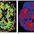
Figure 23-1. Normal myocardial perfusion scintigraphy (MPS). Homogeneous radiotracer uptake in the walls of the left ventricle are shown. A: Apical (left) and basal (right) short axis slices. B: Horizontal long axis. C: Vertical long axis. D: Polar mapping of the same patient demonstrating normal myocardial perfusion.


Interpretation of the Images

Myocardial perfusion and hence tracer distribution is uniform in normal myocardium (Fig. 23-1). A regional defect indicates reduced perfusion in viable myocardium, a reduced amount of viable myocardium, or a combination of both. If a stress defect returns to normal in the resting images, this indicates an inducible perfusion abnormality that is sometimes referred to as inducible ischemia. Strictly, the term “ischemia” is not always correct, particularly if stress is performed with the vasodilators, in which case, the image indicates heterogeneity of perfusion rather than an absolute reduction. Areas of infarction are depicted as a defect in both stress and rest images, and the depth of the defect indicates the amount of myocardial loss. Ischemia can be superimposed upon partial thickness infarction, and in this case, a stress defect may be only partially reversible at rest (Fig. 23-2). Reverse redistribution is the term used to describe a defect in redistribution thallium images that is less apparent in the stress images. When the phenomenon is seen, it is often the result of artifact or other technical differences between images, and, in this case, it is not strictly speaking reverse redistribution. When true reverse redistribution is seen, it is the result of rapid washout of tracer as might occur in an area of partial thickness infarction supplied by a patent artery after angioplasty or thrombolysis. In addition to regional myocardial uptake, other features of the images are important. Ventricular dilation is best judged from the planar images although it is also apparent in the tomograms. Of particular significance is left ventricular dilatation in stress images that is less marked on the resting ones, since this implies extensive inducible ischemia and is associated with an adverse prognosis. The dilatation may be partly real, but it may also be the result of widespread subendocardial ischemia that simulates cavity enlargement. The normal right ventricular myocardium is much thinner than the left, but it is not unusual to see right ventricular uptake. Hypertrophy in either ventricle can be seen as myocardial thickening and an increase in counts. When a relative defect is seen, care must be taken to distinguish between reduced uptake in an area of reduced perfusion and increased uptake in a neighboring area of hypertrophy. Because the myocardial images are not of high spatial resolution, it is often necessary to make this distinction only after echocardiography or MRI has been used to define myocardial thickness.38,39,40 and 41 Increased 201Tl uptake of the lungs is caused by increased pulmonary capillary pressure and is accompanied by an
increased incidence of future cardiac events in patients with CAD. Lung uptake is a marker of impaired left ventricular function at rest or induced by ischemia during exercise. It is expressed as a ratio between uptake in the lungs and the myocardium, even though myocardial uptake depends upon viability and perfusion. The measurements are obtained either from an initial anterior planar image or from the appropriate image of the tomographic acquisition, and a normal lung:heart ratio is less than 0.55 ± 0.11.42 In a mechanism similar to that of 201Tl, the increased uptake of MIBI or tetrofosmin by the lungs and an LHR more than 0.35 ± 0.08 is associated with LV dysfunction and is a predictor of adverse prognosis in patients with known or suspected CAD.43,44 Perfusion defects are not always the result of CAD (see below). Abnormalities can be seen with coronary spasm, anomalous arteries, muscle bridges, small vessel disease as may occur in diabetes or syndrome X, the dilated and hypertrophic cardiomyopathies, hypertrophy caused by outflow obstruction or hypertension, infiltrative disorders such as sarcoidosis and amyloidosis, connective tissue disorders, and conduction defects such as LBBB.


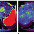
Figure 23-2. Reversible perfusion abnormality in the anteroapical region and the septum superimposed upon minor partial thickness damage at the apex. The perfusion defect is more pronounced towards the apex. The overall appearances are compatible with left anterior descending (LAD) disease. A: Long axis. B: Short axis. C: Polar maps.


Semiquantitative Assessment of Myocardial Tracer Distribution

Several methods have been used to quantify myocardial uptake, and these are all relative rather than absolute techniques in which uptake is expressed relative to the “hottest” voxel (i.e., highest uptake value) within the myocardium. The techniques include linear profiles, circumferential profiles, and analysis of count histograms, but a method that is popular for tomographic imaging is to display the whole myocardium as a polar map or “bull’s-eye” image (Figs. 23-1 and 23-2). Individual polar maps can be compared with a normal database and abnormalities detected automatically as areas with counts less than two standard deviations (or other threshold) from the mean. The normal database should be gender and radionuclide specific and may also be institute specific. The polar mapping is usually visualized with a color scale, where the corresponding pixels below the normal range are displayed with different colors. The extent of myocardial perfusion deficit may be determined as a percentage of the pixels of the polar mapping in which the uptake of the radiopharmaceutical is less than the statistically predetermined threshold. Polar maps with quantification are probably most helpful to assess the depth and extent of defects, but they should not be used as sole basis. However, they can be helpful to assess changes posttreatment. Most experienced nuclear cardiologists prefer to assess the presence of a defect from the tomograms initially and to use quantification to assess depth and extent of an abnormality. The three-dimensional display of LV (3D display) is another option, helping to determine the presence, extent, and location of perfusion defects, while giving information for the size and configuration of LV. Additionally, it can help in correlating SPECT MPI findings with those of other imaging studies, such as those of angiography.7 Segmental analysis can be performed by visual analysis or automated using a number of models of the left ventricular myocardium. A 17-segment model is recommended by several professional societies, and in each segment, tracer uptake is classified semiquantitatively on a five-point scale where 0 = normal (uptake >70%), 1 = mild deficit (uptake 50% to 69%), 2 = moderate deficit (uptake 30% to 49%), 3 = severe deficit (uptake 10% to 29%), and 4 = absence of perfusion (uptake <10%). By adding those numbers together, the sum of the stress scores (SSS), the sum of the rest scores (SRS), or the sum of the difference scores (SDS)45,46 can be calculated (Figs. 23-3 and 23-4).

Because automated analysis has the drawback of being susceptible to artifacts leading sometimes to false-positive results, the findings must not be reported in isolation and without expert review of the images from which the results are derived.7,47 Caution is also required in relating perfusion findings with coronary artery anatomy (Fig. 23-5). Usually, the right feeds the bulk of the inferior wall of the LV and the adjacent portion of the septum. The left after
a short course is divided into the anterior descending artery (LAD) and the circumflex artery (LCX). The LAD feeds the anterior wall and the upper 2/3 of the septum, while the LCX feeds the lateral LV wall and sometimes also the base of the inferior wall. In 85% of cases, the right is the dominant (right dominance), giving rise to the posterior descending artery (PDA). In the rest of the cases, PDA stems from the circumflex artery (left dominance) and perfuses the inferior wall and the lower part of the intraventricular septum or (rarely) from both the LCX and the RCA (codominance).48 Exact matching of anatomical damage and ischemia may be achieved by hybrid imaging, combining CT coronary angiography with SPECT MPI49,50


Figure 23-3. Image interpretation: Assessment of extent and severity of perfusion defects. Short-axis views of four different patients are presented demonstrating a range of perfusion abnormalities (from mild and limited at the left upper corner to extensive and severe at the right lower corner). The extent of a perfusion abnormality is defined by the number of abnormal segments involved, while the severity is determined by the reduction of uptake in each segment. Tracer uptake within a segment is classified as normal; mildly, moderately, or severely reduced; or absent. These categories reflect the counts as a percentage of maximum in the whole set of tomograms. For each category, a score is given, so that SSS, SRS, and SDS can be calculated. Based on the SSS, perfusion images can be classified as normal (SSS 0-3), mildly abnormal (SSS 4-8), or moderate-severely abnormal (SSS ≥ 9).


Figure 23-4. Seventeen-segment model as per American College of Cardiology/American Heart Association (ACC/AHA) recommendations. The left ventricle (LV) has four walls (anterior, septum, inferior, and lateral) and an apex. The LV myocardium can be divided into a certain number of segments, for example, 9, 12, 14, 17 (as it is shown here and recommended by the ACC/AHA), or twenty.


Figure 23-5. Examples of myocardial ischemia (A) of the inferior wall and (B) the lateral wall.


Myocardial Perfusion Studies with Electrocardiographic Synchronization (ECG gated)

During the perfusion study, for assessment of myocardial function, the ECG is recorded while the R wave is used to identify the beginning of the cardiac cycle, as in conventional radionuclide equilibrium ventriculography. This method is used for calculating the ejection fraction (EF) and assessment of segmental contractility disorders of LV (Fig. 23-6). Eight frames per cardiac cycle are commonly used, but sixteen frames per cardiac cycle provide more accurate measurement of left ventricular EF or parameters of diastolic function. By this method, the lower limits of normal left ventricular ejection fraction (LVEF) are calculated around 45% to 50% (in male and female patients, respectively), while regarding the end systolic volume (ESV) and end diastolic volume (EDV), the corresponding values are 45 to 75 and 100 to 150 mL (in female and male patients, respectively).

The segmental movement and systolic thickening are usually studied subjectively, but recent software programs allow the automatic assessment of segmental contractility. Wall motion is best evaluated in linear gray scale without computer-derived edges and can be classified as normal, hypokinetic, akinetic, or dyskinetic (paradoxical). Because of the relatively low spatial resolution of the γ camera, the estimated systolic thickening is done by the periodic variation of scintillations (or counts) between diastole and systole. During the diastolic thinning, a decrease in density of the obtained scintillations is observed, while as the myocardium becomes thickened during systole, the number of scintillations increases. Wall thickening is best evaluated in a continuous color scale without computer-derived edges. Recent software advances permit also assessment of diastolic function and prediction of response to cardiac resynchronization therapy (CRT) using quantitative ECG-gated MPI.51 The timing or phase of contraction of myocardial segments is calculated automatically (phase analysis) and presented in a polar map or phase histogram format. The phase analysis is a parametric image where the regional differences reflect the time difference between the start of contraction of the walls of LV (asynchronous). In normal volunteers, phase analysis values between the RV and LV
are similar while the phase histogram of the ventricles consists of a small amplitude peak. In cases of myocardial ischemia, fibrosis, or conduction disorders, there is enlargement of the histogram.25 The imaging parameters most helpful for predicting response to CRT are histogram bandwidth and phase SD (Fig. 23-7).


Figure 23-6. Myocardial perfusion scintigraphy (MPS) with ECG-gated SPECT. End-diastolic and end-systolic images, representative images of polar mapping, and 3D images of regional wall motion (A). Normal EF and normal end-diastolic and end-systolic volumes (B).


Figure 23-7. A: Example of a patient without LV dyssynchrony on ECG-gated SPECT MPI. Synchronous contraction pattern is reflected by homogeneous phase angle distribution of polar map (left) and narrow highly peaked histogram (right). B: Example of a patient with extensive LV dyssynchrony on ECGgated SPECT MPI. LV dyssynchrony is indicated by heterogeneous phase angle distribution of polar map (left) and wide histogram (right). (Reprinted by permission of the Society of Nuclear Medicine, Boogers MM, Van Kriekinge SD, Henneman MM, et al. Quantitative gated SPECT-derived phase analysis on gated myocardial perfusion SPECT defects left ventricular dyssynchrony and predicts response to cardiac resynchronization therapy. J Nucl Med 2009;50:718-725.)


Assessment of Myocardial Perfusion by Positron Emission Tomography


Positron-Emitting Radiopharmaceuticals

A number of positron-emitting radiopharmaceuticals can be used as tracers of myocardial perfusion. The most common are 82Rb, nitrogen-13 ammonia (13N-ammonia) (Fig. 23-8), and oxygen-15-labeled water (15O-water). 82Rb is a potassium analogue like 201Tl and is the most widely used radiopharmaceutical in clinical practice for MPI with PET. It has a very short physical half-life (76 seconds), but it is produced by a commercially available elution generator with a 4-to 5-week shelf life, thus allowing performance of PET MPI studies in centers without access to cyclotron.52 After an intravenous injection, 82Rb rapidly crosses the capillary membrane and is extracted from plasma by myocardial cells via the Na+/K+ ATPase pump, by an extraction that is similar to that of 201Tl but lower to that of 13N-ammonia and 15O-water. 13N-ammonia is also administered intravenously and has a physical half-life of 9.96 minutes. Once inside the myocyte, 13N-ammonia is incorporated into the glutamine pool and becomes metabolically trapped. The main disadvantage of 13N-ammonia is that it requires an on-site cyclotron and a radiochemistry synthesis capability as well. 15O-water (also a cyclotron produced tracer) can be administered either intravenously or by inhalation of 15CO2 with rapid transformation to water by carbonic anhydrase in the lungs. More recently, a novel MPI tracer has been developed (18F-labeled flurpiridaz). This agent is a structural analogue of pyridaben and binds to mitochondrial complex 1 with high affinity.53 It has a high first-pass extraction fraction of 94% and is currently being evaluated in phase 3 clinical studies. Beyond the tracer kinetics that allows very accurate and reproducible quantification of myocardial perfusion, at least in the experimental setting, the 110-minute half-life of 18F is an additional benefit as it permits a radiopharmaceutical distribution as a singleunit dose on a daily basis. It is estimated that a total administered activity of approximately 520 MBq, for a 2-day protocol, both for stress and rest, provides very satisfactory image quality with acceptable radiation exposure (mean effective dose 0.019 mSv/MBq). For 1-day protocol with adenosine, a minimum dose ratio stress/ rest 2.2 is satisfactory, with a half an hour interval between the two administrations. For 1-day protocol with dynamic exercise, a minimum ratio dose stress/rest 3.0 is required, with a time interval of about 1 hour between the two administrations.54


Stress Tests

Pharmacologic stress is performed using the same agents, as with the SPECT studies. Although pharmacologic stress is the preferred method of inducing hyperemia, exercise testing can also be performed when 13N-ammonia is used as a perfusion tracer (because of the relatively long physical half-life) but is rarely used in practice. In general, both stress and resting injections are administered while the patient is on the scanner’s couch. For assessment of vascular endothelium function, a cold pressor test (CPT) can also be performed, where the patients immerse their left hand in ice water usually for 60 seconds and the tracer is injected while the test continues for another 60 seconds to permit trapping of tracer in the myocardium. CPT is used to assess the health of the vascular endothelium alone as opposed to the stress test performed with the vasodilators, which provides information on the vasomotor function that combines both the endothelial and the smooth muscle cell vasodilator function. CPT has a different action compared to vasodilators as
it is based on sympathetic stimulation. Release of norepinephrine from stimulated sympathetic neurons activates α-adrenoceptors on the endothelium that mediate the release of nitric oxide. In the presence of an intact endothelium, this leads to a 30% to 65% increase in myocardial blood flow (MBF) compared to baseline levels. α-Adrenergic stimulation of vascular smooth muscle cells that causes vasoconstriction is normally counteracted; however, if endothelial integrity is impaired, it prevails over the endothelium-derived vasodilatation. CPT is probably the most appropriate test for early detection of endothelial dysfunction in individuals with risk factors for coronary atherosclerosis, as impairment of the endothelial health precedes abnormalities in smooth muscle cell vasodilator function, but requires extensive procedural standardization.2,51,53


Figure 23-8. Qualitative assessment of myocardial perfusion by stress/rest 13N-ammonia PET. Stress and rest images from a 69-year-old man demonstrating a small area of myocardial infarction at the basal inferior wall and also extensive inducible ischemia involving most of the remaining viable myocardium sparing only the basal septum and the adjacent basal part of the anterior wall. PET, positron emission tomography.


Imaging Protocols and Image Interpretation

Two sets of images are acquired: at rest and following a stress test. In contemporary general purpose scanners that combine PET with CT, PET MPI is accomplished by obtaining a CT scan first for AC followed by perfusion imaging. A scout image or a topogram to determine the precise location of the heart in the thorax and to position the patient correctly is required, and afterward, a low-dose CT scan is performed covering the heart region. This is usually a nongated, fast (<1 minute), low-resolution scan that is obtained during tidal expiration, breath hold, or shallow breathing. Upon termination of this scan, a resting injection of radiotracer is given and resting images are acquired. After completion of the resting images, pharmacologic stress is performed (as for SPECT imaging), during which a second tracer injection is given. A second transmission scan is then acquired for AC. Both rest and stress images can be combined with ECG gating, thus allowing also assessment of global and regional LV function. With 82Rb, total imaging time is less than 30 minutes, while with 13N-ammonia, this takes longer due to the tracer’s longer half-life. The injected activity with 82Rb is 1,500 to 2,500 MBq at rest and the same dose at stress. For 13N-ammonia, the options are either to use a lower activity for rest (370 MBq) and a higher activity for the stress (1,100 MBq) resulting in a faster termination of the study or to inject the same dose (370 MBq) for both studies, but to wait for 13N to sufficiently decay to background levels before the second dose is administered to the patient.2,51,53

Myocardial perfusion and hence tracer distribution is uniform in normal myocardium. It should be noted though that with 13N-ammonia, in normal volunteers, there is a mild heterogeneity in the lateral wall of the LV compared with the other segments. As in SPECT MPI, a reversible perfusion defect indicates myocardial ischemia whilst a fixed defect, myocardial infarction. In addition to regional myocardial uptake, other features of the images are important. Of particular significance is LV dilatation in stress images that is less marked on the resting ones since this implies extensive inducible ischemia and is associated with an adverse prognosis. Enlargement of ventricles seen on both the rest and stress studies indicates ventricular dysfunction. The increased right ventricular (RV) uptake is an indicator of pulmonary hypertension with or without RV hypertrophy. In patients with depressed LV systolic function or chronic pulmonary disease or even in smokers, there might be an increase in the uptake of 13N-ammonia or 82Rb in the lungs. To optimize the contrast between myocardial and background activity, it is better in such cases to increase the time between injection and image acquisition. Attention should be paid also to extracardiac findings in the mediastinum or the lungs observed in the CT scan. These findings might have a clinical importance, as they might be associated with malignancy or an inflammatory process.


As in SPECT MPI, the perfusion defects are characterized qualitatively as mild, moderate, and severe, but also semiquantitatively by obtaining measurements (scores) of segmental perfusion according to the standard of 17 segments (see above).


Quantification

In human studies, PET is the gold standard for quantifying myocardial perfusion in absolute terms (mL/min/g) both at rest and stress and also the assessment of coronary vasodilator reserve (CFR). This is because PET has a high sensitivity and temporal resolution, which allow fast dynamic imaging of tracer kinetics and extensively validated algorithms for accurate correction of photon attenuation. Global and regional myocardial perfusion is measured by compartmental (usually one or two tissue compartments) tracer kinetic models, which are then combined with appropriate corrections for physical decay of the radioisotope, partial volume-related underestimation of the true myocardial tissue concentrations, and spillover of radioactivity between the left ventricular blood pool and the myocardium.15O-water is the ideal tracer for quantification as it diffuses freely across plasma membranes and exhibits a linear relationship between uptake and flow at high flow rates. However, other PET radiopharmaceuticals are quite satisfactory for quantification as well.51,53 82Rb compares reasonably well with 13N-ammonia or 15O-water in absolute measurements of myocardial perfusion, and it is therefore an attractive option for hospitals without easy access to a cyclotron.


▪ Clinical Applications of Radionuclide Myocardial Perfusion Imaging


The Role of SPECT MPI in the Diagnosis of CAD

The hallmark of stable CAD is the predictable central chest pain provoked by exertion and relieved by rest. This is caused by obstructive atherosclerosis that results in reduction of myocardial perfusion (and thus ischemia) upon exhaustion of CFR. The need for and the appropriate choice of further investigation of chest pain should be decided on the basis of the pretest likelihood of significant CAD, which can be estimated by different predictive nomograms. Amongst them, the Diamond and Forrester predictive table integrates three clinical variables (quality of chest pain, gender, and age) to provide an estimation of the likelihood of angiographically significant coronary disease.55 This approach can be refined by incorporating other powerful predictors, such as serum cholesterol levels, systolic blood pressure, and diabetes.56 Most predictors of pretest likelihood are able to distinguish those at low and high likelihood of CAD but leave many to be classified as intermediate. According to Bayes’ theorem,57 these patients have the most to gain from further investigation, as a positive or negative result allows reclassification of intermediate pretest likelihood to either high or low posttest likelihood, respectively. A positive or negative result can revise the intermediate value to high or low, respectively, while a positive test in patients with low probability of CAD (<15%) is likely to be a false positive, and thus, further investigation would only increase the cost, exposing the patient to unnecessary procedures and risks. On the other hand, most patients with high pretest probability of disease (>85%) do not require further noninvasive investigation for diagnostic purposes, but functional imaging can be performed for risk stratification and management decisions. Exercise electrocardiography (ExECG) remains the test of first choice in many centers, especially in the United States, because it is widely available, is easy to perform, and does not involve radiation exposure. Noninvasive CT coronary angiography is performed in patients unable to exercise or when a stress test is contraindicated, but in some centers, it is a first-line investigation in patients with low to intermediate likelihood of CAD, because it can be carried out quickly and easily and has a high negative predictive value (NPV) (up to 98%).58 It should be pointed out that while the latter provides information on the morphology of the lumen of the coronary vessels and whether there is stenosis, a stress test enables assessment of hemodynamic response, ECG changes, symptoms, and exercise tolerance during stress, which probably match (or in several instances exceed) the usual level of a patient’s activity. Meta-analyses indicate that the average sensitivity and specificity of ExECG are 68% and 77%, respectively.59 ExECG is of limited value if the patient cannot exercise adequately or has an abnormal resting ECG that precludes ST-segment assessment on exercise (preexcitation, left ventricular hypertrophy, and changes due to the use of digoxin). In these patients, but also in those with a nondiagnostic ExECG result, stress/rest MPI is ideally suited for use in the diagnosis of stable CAD due to its ability to assess directly myocardial perfusion and its alterations during exercise or pharmacologic stress. Normal myocardial perfusion after stress indicates the absence of functionally significant CAD and is associated with a low risk (<1%; see below) of future coronary events.60

It should be noted that normal stress perfusion is not synonymous with the absence of CAD, as coronary atherosclerosis need not necessarily cause stenosis of sufficient severity to result in impairment of CFR. In the presence of abnormal perfusion, the site, depth, and extent of the demonstrated ischemia provide diagnostic information and can guide subsequent management. On a recent meta-analysis, sensitivity and specificity were 85% to 90% and 70% to 75%, respectively, for the detection of angiographically significant CAD.61 It is important to recognize that conventional coronary angiography (ICA) is an anatomical rather than a functional technique and that a “significant” stenosis on ICA may be associated with normal CFR. In clinical practice therefore, and unless ICA is combined with functional indexes of the coronary status (e.g., intracoronary Doppler velocity wire flow reserve or coronary pressure wire measurements), the techniques should be seen as providing complementary rather than necessarily equivalent information.

The sensitivity and specificity of SPECT MPI can be improved using ECG gated62 and AC. ECG gated allows quantitation of global and regional ventricular function that, aside from providing important prognostic information, aids in the distinction of artifacts from true perfusion defects, thus improving specificity and normalcy. Fixed defects in the anterior and inferior walls, commonly due to breast and diaphragmatic attenuation artifacts in women and men, respectively, may be mistaken for myocardial infarction, but myocardial motion and thickening in these areas are normal on gated images. Similarly, the use of AC to recover counts in these areas further improves the recognition of attenuation artifacts and hence diagnostic accuracy. A common cause of suboptimal specificity of SPECT MPI reported in earlier studies is posttest referral bias. In other words, when the reference standard (in this case ICA) is not performed in all patients and referral to angiography is more likely when SPECT MPI is reported as abnormal, then the results are affected by posttest referral bias and specificity appears falsely low. Normalcy, the number of patients at low likelihood of coronary disease with normal SPECT MPI, is a better index for assessing the performance of the test for excluding disease, and this was found to be consistently high (around 90%) irrespective of tracer or method of stress.61


SPECT MPI in Risk Stratification and Management of Patients


Risk Assessment in Patients with Suspected or Confirmed CAD

The risk of future cardiac events, such as myocardial infarction or death, is a significant determinant of treatment type. A number of factors, such as extent of anatomic disease, total ischemic burden, degree of LV dysfunction, threshold of angina, presence of
comorbidities such as severe peripheral vascular disease or diabetes, and uncontrolled CAD risk factors, contribute to the annual risk of cardiac events, which may be classified as low (<1%), intermediate (1% to 3%), or high (>3%).63 Numerous studies have confirmed the excellent prognostic power of SPECT MPI and its important role in risk stratification and patients’ management.64,65 and 66 In addition, SPECT MPI provides incremental information over and above that obtained by clinical or stress ECG data. Several large studies reporting prognostic analyses of MPI in cohorts using exercise, vasodilator, or both types of stress in various clinical settings have documented the incremental value of MPI.67,68 and 69 The incremental value of SPECT MPI can be increased further for assessing the probability of death due to cardiovascular causes by incorporating information on LVEF and LV end-systolic volume derived from the gated data.

A normal SPECT MPI is associated with a 0.7% annual risk of MI and cardiac death, which is similar to the general population.70 This has important clinical implications because a normal study obviates the need for invasive investigation or revascularization. It should be noted, however, that the value 0.7% per year represents an average annual event rate and a normal perfusion study should always be interpreted in the context of the individual patient. For instance, patients with normal myocardial perfusion but significant ST segment depression during adenosine stress are at increased risk for nonfatal myocardial infarction, (7.6% compared to 0.5% in patients without such findings, during a mean follow-up period of 29 +/- 12 months).71 In addition, higher heart rate at rest and a blunted heart rate increase during adenosine infusion have also been found to be predictors of severe future events. Specific groups, such as the elderly, those with diabetes, or those with known CAD, have an annual event rate somewhat higher (1.4% to 1.8% per year) despite normal SPECT MPI.65 In general, the “warranty period” of a normal SPECT MPI in this setting is around 2 years, after which repeat scanning may be necessary to redefine prognosis.

An abnormal scan confers around a sevenfold increase in annual cardiac events compared to a normal study.72 The probability of a cardiovascular event increases in parallel with increasing depth and extent of reversibility of inducible ischemia, which are markers of the stenosis severity and the amount of myocardium subtended by the stenosed vessels, respectively. Those with only mild inducible ischemia have an annual event rate of around 3%, which rises to 7% in those with severe ischemia. The presence of corollary markers of severe 3-vessel disease, such as transient left ventricular dilatation73 or increased lung uptake of 201Tl,74 increases the event rate still further (Table 23.4).


TABLE 23.4 High-Risk Imaging Variables


Multiple myocardial perfusion defects


Extensive reversibility of a perfusion defect


Transient left ventricular dilation


Multiple regional wall motion or wall thickening abnormalities


Left ventricular ejection fraction <35%


Increased end-diastolic and/or end-systolic volume in ECG-gated SPECT


Increased lung tracer uptake


LV dyssynchrony assessed by phase analysis


Patients with mildly abnormal SPECT MPI are generally at low risk of cardiac death; therefore, if they are not limited by their symptoms, these patients might be candidates for aggressive medical therapy/risk factor modification. However, this is not the case in patients with significant comorbidities (e.g., advanced age, prior CAD, diabetes mellitus, atrial fibrillation, pharmacologic stress, etc).68 The decision, therefore, whether or not to catheterize a patient with a mildly abnormal perfusion scan becomes a function of the underlying patient condition.

In the current era, SPECT MPI is used increasingly to test the effectiveness of different management strategies both on a clinical basis and cost-effectiveness basis. The largest and most representative study included 13, 969 patients who were investigated with SPECT MPI and followed up over 8 years.75 The results showed that patients without extensive scar but significant (at least 10%) ischemic myocardium were likely to realize a survival benefit from early revascularization. In contrast, patients with minimal ischemia fared better on medical therapy without early revascularization. These findings agree with those from the SPECT MPI substudy of the Clinical Outcomes Using Revascularization and Aggressive Drug Evaluation trial, which included 314 patients with known CAD.76 Patients who were randomized to receive percutaneous coronary intervention (PCI) in addition to optimal medical therapy (OMT) had a greater reduction in inducible ischemia than did those randomized to receive OMT alone. Reduction in ischemia was associated with improvement in angina frequency and stability, and this was more pronounced in patients with baseline moderate-to-severe ischemia.

Only gold members can continue reading. Log In or Register to continue

Stay updated, free articles. Join our Telegram channel

Jul 8, 2020 | Posted by in ULTRASONOGRAPHY | Comments Off on Myocardial Perfusion Imaging with PET/SPECT: Techniques and Clinical Applications

Full access? Get Clinical Tree

Get Clinical Tree app for offline access