Figure 26.1.1
Intramural fibroid. A: Sagittal (SAG UT) and (B) coronal (COR UT) transvaginal views of the uterus demonstrate a large fibroid (calipers) within the body of the uterus. C: Color Doppler image in coronal plane demonstrates considerable blood flow around and within the fibroid, indicating that it is highly vascular.
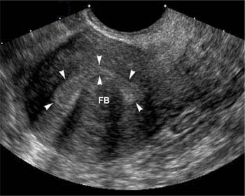
Figure 26.1.2
Submucosal fibroid. Sagittal view of the uterus demonstrates a submucosal fibroid (FB) indenting the endometrium (arrowheads).
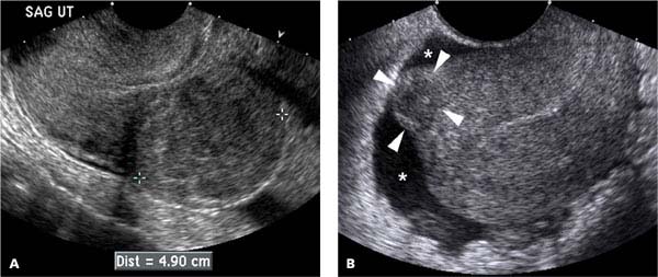
Figure 26.1.3
Subserosal fibroids. Sagittal transvaginal views of the uterus in two patients demonstrating (A) a large subserosal fibroid (calipers) extending outward from the posterior wall of the uterus; and (B) a small subserosal fibroid (arrowheads), surrounded by free fluid (*) in the pelvis, extending outward from the anterior fundal aspect of the uterus.
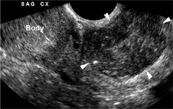
Figure 26.1.4
Cervical fibroid. Sagittal view of the cervix (SAG CX) demonstrates a large cervical fibroid (arrowheads) located caudal to the uterine body (Body).
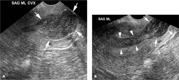
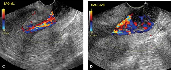
Figure 26.1.5
Fibroid prolapsing into the cervix. A: Sagittal midline view of the cervix (SAG ML CVX) demonstrating a hypoechoic mass (arrows) within the cervix. B: Sagittal midline view of the uterus shows that the cervical mass (arrows) extends from the body of the uterus via a stalk (arrowheads). C and D: Color Doppler documents that the fibroid in the cervix is attached to the stalk, by showing continuity of blood vessels between these two structures.
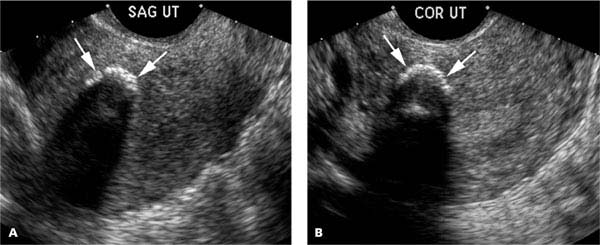
Figure 26.1.6
Calcified fibroid. A: Sagittal (SAG UT) and (B) coronal (COR UT) transvaginal views of the uterus demonstrate a rim of calcification (arrows) with posterior acoustic shadowing, representing calcification in the wall of a fibroid.
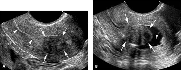
Figure 26.1.7
Saline infusion sonohysterogram of a submucosal fibroid. A: Sagittal transvaginal view of the uterus demonstrates a fibroid (arrows) abutting the endometrium (arrowheads). B: After instillation of saline, the fibroid (arrows) is seen projecting into the fluid-filled (F) uterine cavity.
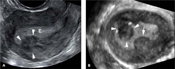
Figure 26.1.8
3D ultrasound of a submucosal fibroid. A: Sagittal transvaginal view of the uterus demonstrates a fibroid (arrowheads) projecting into the endometrium (E). The dotted green line represents the line perpendicular to which a multiplanar reconstruction from 3D ultrasound was performed. B: The image in the reconstructed plane shows that the fibroid (arrowheads) projects more extensively into the endometrium than was evident on the prior image, and also demonstrates a second submucosal fibroid (arrow).
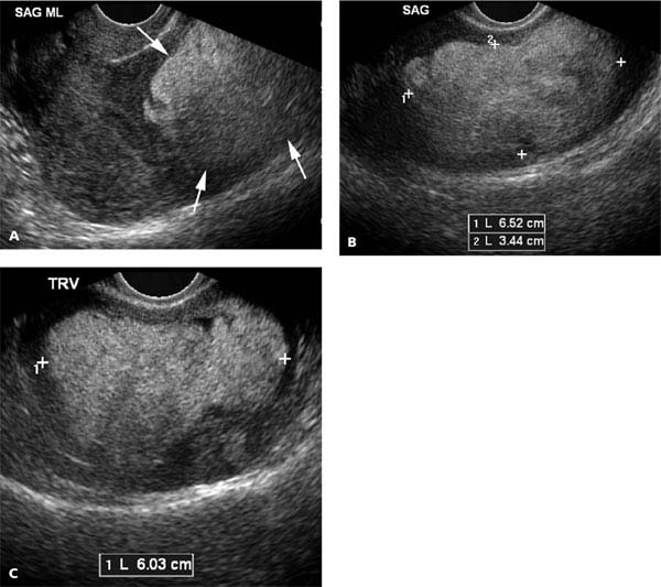
Figure 26.1.9
Lipoleiomyoma. A: Sagittal midline (SAG ML) view of the uterus demonstrates a large echogenic mass (arrows) in the body and fundus of the uterus. The echogenic nature of the mass indicates that it has a high fatty content, consistent with lipoleiomyoma. B: Sagittal (SAG) and (C) transverse (TRV) view better demonstrate the borders and size of the lipoleiomyoma (calipers).
Lipoleiomyomas appear as echogenic masses within the uterus (Figure 26.1.9). The echogenic appearance is due to the adipose tissue in the mass.
Leiomyosarcomas have a sonographic appearance similar to that of fibroids. Because they are much less common than fibroids, they are not generally diagnosed preoperatively. If a mass that appears to be a fibroid enlarges on serial sonograms in a postmenopausal woman, the diagnosis of leiomyosarcoma should be suspected (Figure 26.1.10).
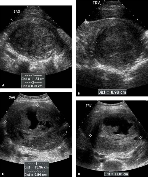
Figure 26.1.10
Leiomyosarcoma. A: Sagittal (SAG) and (B) transverse (TRV) transabdominal views of the uterus in a postmenopausal woman demonstrate a hypoechoic mass (calipers) in the uterus. This was initially diagnosed as a fibroid. C: Sagittal and (D) transverse views 5 months later show that the mass has grown substantially. In view of the increasing size of the mass in a postmenopausal woman, leiomyosarcoma was felt to be the likely diagnosis. Hysterectomy was performed, and the diagnosis of leiomyosarcoma was confirmed by pathology.
Related posts:


Stay updated, free articles. Join our Telegram channel

Full access? Get Clinical Tree


