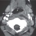
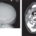
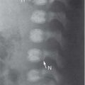
Fig. 4.135 Occipitomental radiograph shows right nasal radiopaque foreign body.Fig. 4.134 Axial CT (bone window) shows left posterior choanal atresia.Fig. 4.136 Lateral radiograph of the nasopharynx shows a soft-tissue mass projected over the nasopharynx representing hypertrophy of nasal turbinate.Fig. 4.137 Coronal CT (bone window) shows bilateral nasal, maxillary, and ethmoid sinus opacification in polyposis.Fig. 4.138 Coronal CT (bone window) shows a well-circumscribed, expansile osseous lesion with a central nonossified portion centered in the left ethmoid sinus and nasal passage, which has the characteristic appearance of an ossifying fibroma.
Only gold members can continue reading. Log In or Register to continue