Massive mural thickening of cecal ± ascending colon wall
– Other segments of colon and small bowel can be affected
Mucosal hyperenhancement and submucosal edema (marked)
Infiltration of pericolonic fat
• Less common, more severe findings
Pneumatosis, extraluminal gas and fluid (perforation)
TOP DIFFERENTIAL DIAGNOSES
• Pseudomembranous colitis
• Cecal diverticulitis
• Crohn colitis
PATHOLOGY
• Severely neutropenic patients
• Majority of cases are those with leukemia &/or hematopoietic stem cell transplant recipients
• Pathogenesis: Probably due to combination of factors
Mucosal injury by cytotoxic drugs
Profound immunosuppression
Invasion of bowel wall by microorganisms (polymicrobial)
Progressive necrosis of bowel wall
CLINICAL ISSUES
• Fever, RLQ tenderness in immunosuppressed patient
• Watery diarrhea, ± hematochezia
DIAGNOSTIC CHECKLIST
• Consider history of chemotherapy for leukemia or bone marrow transplantation
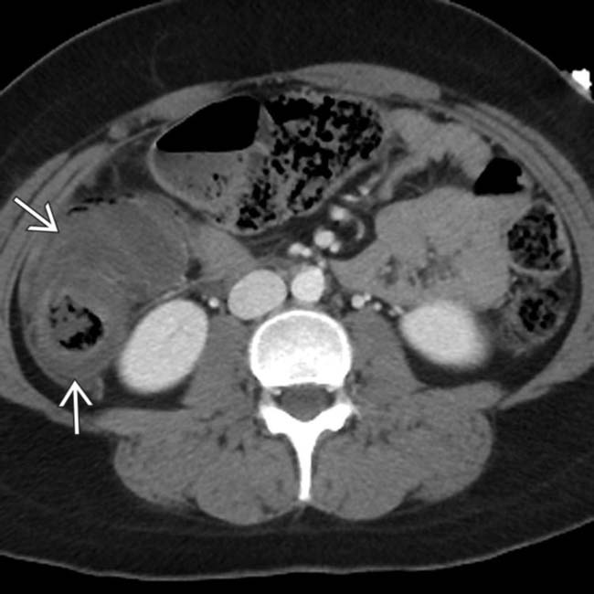
(Left) This 25-year-old woman was receiving chemotherapy for synovial sarcoma and became severely neutropenic, with complaints of abdominal pain, fever, and diarrhea. Axial CECT shows marked submucosal edema of the cecum and ascending colon .
(Right) Another CT section in the same patient shows mucosal hyperenhancement and submucosal edema.
(Left) Coronal reformatted CT in the same patient shows inflammation of the ascending colon and terminal ileum , along with pericolonic ascites .
(Right) Another coronal CT section in the same case shows pneumatosis within the cecal wall, which, along with ascites , suggested perforation, subsequently proven at surgery. Necrotizing, neutropenic colitis was the diagnosis.
TERMINOLOGY
Synonyms
• Neutropenic enterocolitis, ileocecal syndrome
Definitions
• Life-threatening, necrotizing enterocolitis occurring primarily in severely neutropenic patients
IMAGING
General Features
• Best diagnostic clue
Massive mural thickening of cecal ± ascending colon wall
• Location
Cecum, ascending colon ± distal ileum
– Other segments of colon and small bowel can be affected
• Morphology
Dilated/narrowed lumen, thickened wall
Radiographic Findings
• Radiography
Ileocecal dilatation with air-fluid levels
“Thumbprinting” in wall of ascending colon, ± pneumatosis
CT Findings
• NECT
Circumferential wall thickening of cecum ± ascending colon and distal ileum
Decreased bowel wall attenuation due to edema
Only gold members can continue reading. Log In or Register to continue


 .
.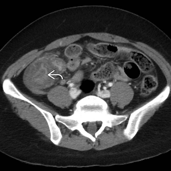
 and submucosal edema.
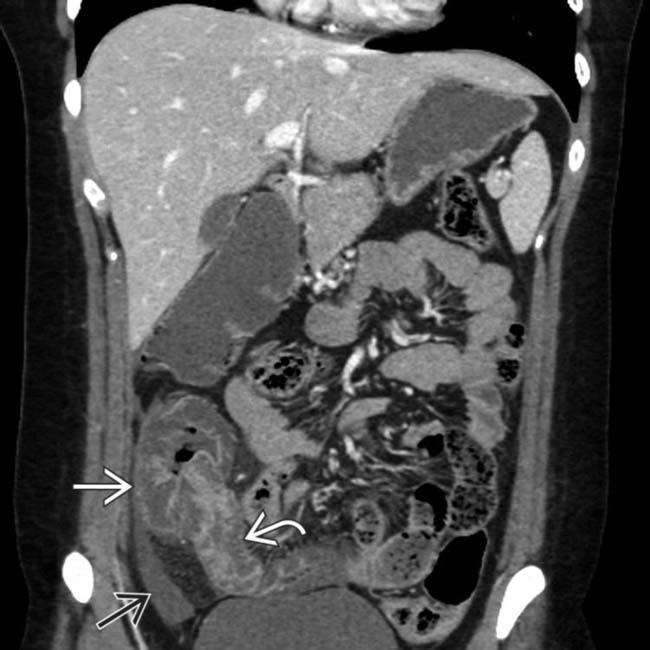
 and terminal ileum
  , along with pericolonic ascites
  .
.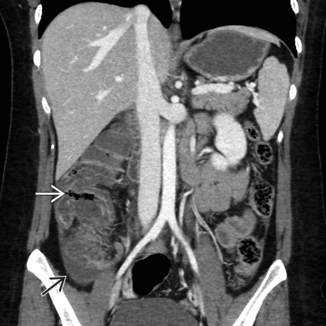
 within the cecal wall, which, along with ascites
  , suggested perforation, subsequently proven at surgery. Necrotizing, neutropenic colitis was the diagnosis.


