Axis
Transverse process
Iliac wing
Sacral ala
7 cervical vertebral bodies
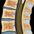
12 thoracic vertebral bodies
5 lumbar vertebral bodies
5 fused sacral vertebral bodies
4 coccygeal bodies
(Top) Coronal graphic of the spinal column shows relationship of 7 cervical, 12 thoracic, 5 lumbar, 5 fused sacral, and 4 coccygeal bodies. Note cervical bodies are smaller with neural foramina oriented at 45° and capped by the unique C1 and C2 morphology. Thoracic bodies are heart-shaped, with thinner intervertebral discs, and are stabilized by the rib cage. Lumbar bodies are more massive, with prominent transverse processes and thick intervertebral discs.
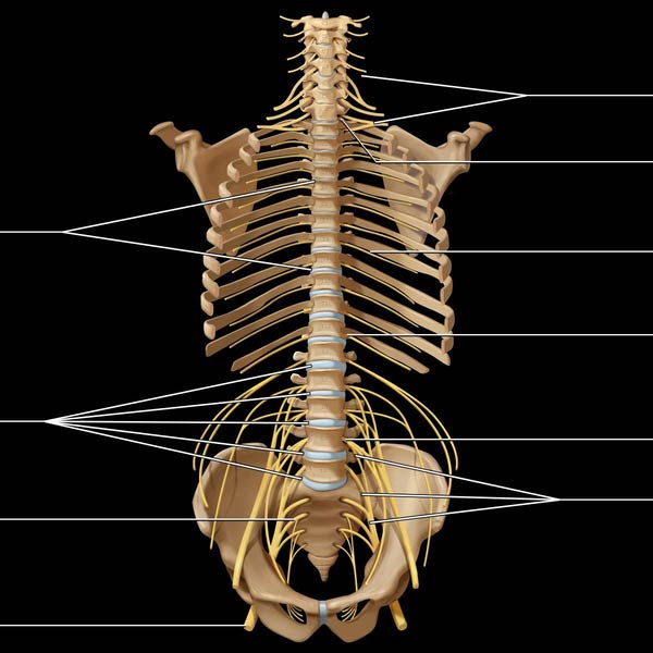
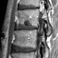
Lumbar intervertebral discs
Sacral nerve roots
Sciatic nerve
Brachial plexus
C8 root exiting at C7-T1 level
Intercostal nerves
T12 root exiting at T12-L1 level
L4 root exiting at L4-L5 level
Lumbosacral plexus
(Bottom) Coronal graphic demonstrates exiting spinal nerve roots. C1 exits between the occiput and C1, while the C8 root exits at the C7-T1 level. Thoracic and lumbar roots exit below their respective pedicles.
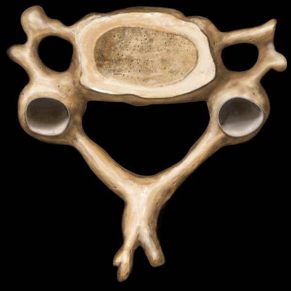
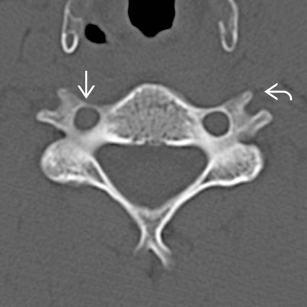
 , encompassing the vertical course of vertebral artery. The anterior and posterior tubercles
  give rise to muscle attachments in neck.
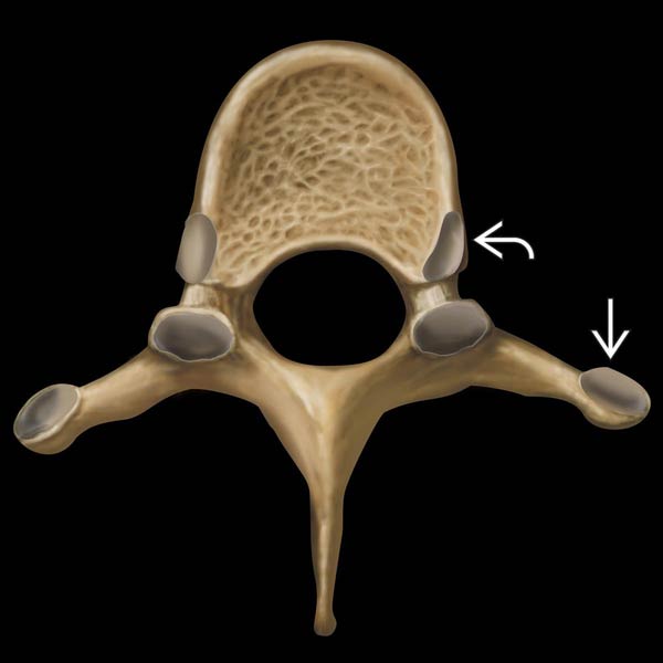
 and costovertebral joints
  . Facet joints are oriented in the coronal plane.
Related posts:

Stay updated, free articles. Join our Telegram channel

Full access? Get Clinical Tree


