Normal Growth, Normal Development, and Congenital Disorders


Normal Growth, Normal Development, and Congenital Disorders


Victor Ho-Fung

Adji Saptogino

Timothy Cain

Karuna M. Das

Selim Doganay

Diego Jaramillo


INTRODUCTION

The understanding of normal growth and development of the musculoskeletal system is fundamental for the evaluation of the wide spectrum of congenital and developmental abnormalities in children. This knowledge is essential for accurate identification of imaging findings, differential diagnosis, classification, and therapeutic guidance. In this chapter, the currently available various imaging modalities and their main uses in pediatric patients are discussed. The normal anatomy of the developing skeleton is reviewed. An overview of the important imaging characteristics of skeletal abnormalities and syndromes related to skeletal growth and development is provided. In addition, a concise description of the underlying pathophysiology, imaging diagnosis, and therapeutic approaches are also presented.


IMAGING TECHNIQUES


Radiography

Conventional radiographs remain the principal imaging modality for the diagnosis of skeletal abnormalities and skeletal dysplasias. The evaluation of congenital and developmental abnormalities in neonates and infants is based upon the identification of specific morphologic patterns in the different components of the developing skeleton. In the case of skeletal dysplasia, both axial and appendicular skeletal features require a complete assessment using skeletal survey protocols. There are useful comprehensive textbooks dedicated to the evaluation of skeletal dysplasias1 and anatomic variants.2


Ultrasound

The role of ultrasound (US) in musculoskeletal imaging is currently expanding beyond the evaluation of the infant hips. The lack of ionizing radiation exposure or need for sedation, wide availability of US equipment, and the advantage of tailored examinations with both static and dynamic components are all strengths of US, in particular for neonates and young children with abundant hyaline cartilage. Prenatal US is the primary method for evaluation of the fetus and is not unusual to first suspect a potential skeletal dysplasia during routine US examination following identification of shortened long bones or other abnormal skeletal findings.3,4 However, the accurate prenatal diagnosis of skeletal dysplasias remains challenging because of the heterogeneity of this group of relatively rare disorders, variability in the time of manifestation of clinical findings, and frequent lack of a corroborating genetic and molecular diagnosis.5,6,7 A multidisciplinary approach among radiologists, clinicians, pathologists, and geneticists is crucial for the correct diagnosis of skeletal dysplasias and to maximize the usefulness of genetic counseling to the parents.


Computed Tomography

Computer tomography (CT) with multiplanar and three-dimensional (3D) reformation is an ancillary tool to conventional radiography for assessment of skeletal abnormalities, particularly during surgical planning as in the case of tarsal coalitions and transitional fractures.8,9 The risks of radiation exposure versus the clinical benefits of CT examinations should always be carefully considered in the pediatric population.10,11 In the case of the long bones in the extremities,
potential radiation risks are lower than in the axial skeleton, which is located near more radiosensitive mediastinal, abdominal, and pelvic organs.12 More recently, selective use of fetal CT with 3D reformations has been described in cases in which US and genetic data are inconclusive to either diagnose or exclude a suspected skeletal dysplasia with impact on accurate counseling of families.5,12,13,14,15


Magnetic Resonance Imaging

Magnetic resonance imaging (MRI) allows comprehensive evaluation of bone marrow, cartilage, and soft tissues in children with its inherent superior contrast resolution. The capacity to confidently identify different types of hyaline cartilage (physeal, epiphyseal, articular) and their abnormalities in growing children is one of the main advantages of MRI.16

The bone marrow is the main site for hematopoiesis. The two normal types of bone marrow consist of red (hematopoietic marrow) and yellow (fatty marrow). The cellular composition of normal marrow changes in an orderly fashion with age (from red to yellow marrow), a process known as marrow conversion (Fig. 21.1).

MRI allows for identification of expected age-related changes through the skeleton. At birth, hematopoietic marrow is present in the entire skeleton. Subsequently, marrow conversion begins in the periphery of the skeleton. This occurs first along distal phalanges (fingers and toes), in a symmetric, centripetal distribution into the axial skeleton. Following the appearance of the secondary center of ossification, both epiphyseal and apophyseal regions are the first to developed fatty marrow within months of ossification (showing signal characteristics of fat tissue on both T1 and fluid-sensitive sequences), an important observation when evaluating marrow signal intensity on infants and young children. In the long bones, marrow conversion occurs during the first decade of life. In these areas, the process begins along the diaphysis and progresses to the distal metaphysis and finally into the proximal metaphysis. Small residual areas of hematopoietic marrow with typical straight margins or feathery-shape appearance can be seen in the metaphyseal regions, particularly in the proximal femur of adolescents. Persistence of large amounts of hematopoietic marrow in the diaphysis after 10 years of age is considered abnormal. The marrow conversion process continues into the mid to late third decade of life, when bone marrow distribution reaches adult state (Fig. 21.1).17


FIGURE 21.1 Normal bone marrow conversion in long bones. Sequential age-related changes in hematopoietic marrow to fatty marrow distribution are illustrated in this figure representing a femur.

Multiple factors requiring increased hematopoiesis can cause marrow reconversion (from yellow to red marrow) such as anemias (sickle cell and thalassemia), physiologic stress (high altitude, endurance training, obesity), and chemotherapy with granulocyte colony-stimulating factor therapy. Also, abnormal marrow replacement can be seen with neoplastic disease (leukemia and metastatic disease).

Potential limitations of MRI include cost, availability, and the need for sedation in younger children. However, the role of MRI for prenatal characterization of skeletal dysplasia is limited, primarily because of difficulty in displaying a panoramic image of a fetus and obtaining adequate images for visualization of the main axis of individual bones.13


Nuclear Medicine

Nuclear medicine is a useful imaging modality for specific skeletal dysplasias related to metabolically active bone and soft tissue lesions. For example, the use of bone scintigraphy has been described for identification of bone lesions in polyostotic fibrous dysplasia.18 Also, the role of hybrid imaging combining positron emission tomography (PET) functional imaging with the anatomic detail of CT (PET-CT) in children is expanding, particularly in the characterization and staging of neoplastic disease.19,20,21


NORMAL ANATOMY

The skeletal system develops in utero from a condensation of primitive mesenchymal cells that are precursors of cartilage or membranous bone. Membranous ossification refers to bone formation through mesenchymal differentiation directly into osteoblasts without a cartilaginous precursor. Membranous ossification is responsible for the development of the facial bones and cranium. In the long bones, the periosteal membrane envelopes the outer layer of the bone and is responsible for membranous ossification resulting in axial growth.

The structure of the tubular bones is similar through the body. A synchondrosis refers to a joint in which the connecting medium is hyaline cartilage, such as the epiphysis of long bones and sternocostal joints. The epiphyses are at both ends of most long bones, with each epiphysis located between the joint and the primary physis. The epiphyses are initially completely cartilaginous, followed by the
development of a secondary ossification center and progressive conversion into bone.

Endochondral ossification refers to bone formation through a cartilaginous model. It is responsible for the development of the skull base, long bones, clavicles, and vertebral column. In the long bones, endochondral ossification allows longitudinal growth with formation of columns of cartilage in the primary physis with sequential morphologic changes and transformation of the chondrocytes in new bone at the metaphysis.22

The apophyses do not contribute to the longitudinal growth of long bones but provide important structural support to the insertion of muscles, tendons, and ligaments. The apophyseal formation is similar to the epiphyses; it is initially entirely cartilaginous with subsequent conversion into bone because of the development of a secondary ossification center and a physeal equivalent.

The epiphyseal cartilage is a vascularized structure supplied by vascular canals. The epiphyseal vascular canals are composed of nonanastomotic arterioles, venules, and sinusoidal capillaries that are radially arranged around the secondary center of ossification.23 The physeal cartilage is only vascular during the first 18 months of life and then becomes avascular. These changes also determine the pathologic changes seen at different stages of skeletal maturation. For example, vascular canals are prominent in diseases that result in inflamed synovium, such as juvenile idiopathic arthritis, and when infection invades the epiphyseal cartilage. They are attenuated when there is ischemia of the cartilage, such as that resulting from excessive abduction in spica casts for developmental hip dysplasia treatment or that related to large-joint septic arthritis.

The metaphysis is the most vascularized structure of the growing skeleton. Systemic processes are manifested first in these regions along the appendicular skeleton, in particular in rapidly growing bones such as distal femur and proximal tibia. The interaction of the metaphysis on the adjacent physis allows formation of new bone from the cartilaginous template of the physis. Both physiologic and pathologic processes can have similar appearance along the metaphyseal regions, and knowledge of these variants is important for the imaging evaluation in children.

The differential diagnosis for dense metaphyseal transverse bands is extensive, but among the most important considerations are normal dense zones of provisional calcification (most common in neonates), treated leukemia, chronic lead poisoning, hypoparathyroidism, healing rickets, scurvy, and “growth recovery lines” (seen in patients with generalized systemic disease causing periods of chronic illness and recovery).

The differential diagnosis for lucent metaphyseal transverse bands includes normal variation, leukemia and metastatic neuroblastoma, and TORCH (toxoplasmosis, rubella, cytomegalovirus, herpes simplex, and HIV) infection in the neonate. The presence of perpendicular linear bands of metaphyseal lucency and sclerosis (striations) can be seen as a normal variant along the proximal femurs.


DEVELOPMENTAL AND ANATOMIC VARIANTS


Metaphyseal Beaking and Genu Varum

Metaphyseal beaking, particularly in the medial aspect of the proximal tibia, and lower extremity bowing (genu varum) are not uncommon in young children. Genu varum usually is seen in walking children under 2 years of age with physiologic resolution after this age (Fig. 21.2 and Schematic N).24 The differential diagnosis for pathologic genu varum includes trauma, skeletal dysplasia (achondroplasia), rickets, fibrous dysplasia, and Blount disease. Metaphyseal fragmentation with physiologic bowing has also been described in young children and considered physiologic in children after ˜18 months of age.25


Physiologic Periosteal Reaction

Physiologic periosteal reaction is an important developmental variant in young infants between 1 and 6 months. The typical features of physiologic reaction include smooth, thin (<2 mm), and symmetric distribution along rapidly growing long bones, particularly the humeri, femurs, and tibias (Fig. 21.3). The periosteum, which is responsible for the membranous ossification

of long bones, also guides the reparative response to stimuli (e.g., trauma, infection, neoplasm, metabolic disorder, and nutritional status). In children, the periosteum is physiologically more active and less adherent to the cortex than in adults, allowing for earlier and more aggressive appearance of the periosteal reaction.26 The differential diagnosis for benign periosteal reaction in the pediatric population is summarized in Table 21.1.


FIGURE 21.2 Physiologic genu varum and metaphyseal beaking in a 19-month-old girl. Frontal radiograph of bilateral lower extremities shows symmetric genu varum with medial metaphyseal beaking in both distal femurs (arrows) and proximal tibias (interrupted arrows). Patient was asymptomatic and genu varum resolved 1 year after radiographic evaluation.


image


FIGURE 21.3 Physiologic periosteal reaction in a 2-month-old boy. A: Frontal radiograph of the right tibia. B: Frontal radiograph of the left tibia. Both images show thin, smooth, continuous, symmetric periosteal reaction (arrows) along the medial diaphyseal cortex of both tibias. Included portions of the distal femurs also show physiologic periosteal reaction (interrupted arrows).


Cortical Desmoid

A very common developmental variant in older children is a cortical desmoid (also known as distal femoral cortical irregularity) (Fig. 21.4 and Schematic L). This lesion is a self-limiting fibrous or fibroosseous lesion most commonly located in the medial supracondylar region, believed to be a tug lesion at the insertion of the adductor magnus aponeurosis or the origin of the medial head of the gastrocnemius.27,28 Prevalence is highest in boys age 10 to 15 years. The majority of affected pediatric patients are asymptomatic, although some have a history of knee pain secondary to trauma.


TABLE 21.1 Differential Diagnosis of Physiologic Periosteal Reaction (1-6 Months of Age)


Healing fracture


Infection


Drugs (prostaglandins, hypervitaminosis A)


Hypertrophic osteoarthropathy (cardiopulmonary diseases, malignancy)


Infantile cortical hyperostosis (Caffey disease)


Radiographic evaluation typically demonstrates a cortical irregularity or erosion with a sclerotic base at the posteromedial distal femur in the lateral view and occasionally a well-circumscribed lucent region with a sclerotic thin rim in the frontal view. MRI typically demonstrates a cortical irregularity along the posteromedial distal femur with increased T2 signal intensity within the adjacent bone marrow. Occasionally, associated traumatic periostitis and swelling with soft tissue edema at the origin of the medial head of the gastrocnemius can be seen.27 Typical radiographic features are diagnostic for this entity, and in the presence of a benign clinical course, further workup and biopsy can be avoided.29


Discoid Meniscus

Discoid meniscus is a congenital anatomic variant seen more commonly in the lateral meniscus (Fig. 21.5 and Schematic M). The incidence varies from 0.4% to 16.6% on arthroscopic studies.30 A discoid meniscus is more disc shaped than semilunar shaped in configuration with a portion of the meniscus extending to the central portion of the tibial plateau.31 The majority of affected pediatric patients are asymptomatic; however, the abnormal configuration of the discoid meniscus alters biomechanics and causes a predisposition for increased meniscal tears and intrasubstance degeneration. Symptomatic pediatric patients present with a “snapping knee” and, when torn, can be a cause of locking and knee pain.


FIGURE 21.4 Cortical desmoid in a 4-year-old boy. A: Frontal radiograph of the right knee shows a lucent lesion (arrow) with well-marginated sclerotic borders in the medial femoral metadiaphysis. B: Lateral radiograph of the right knee shows the typical posterior location with sclerosis and mild irregularity (arrow). C: Axial fat-saturated T2-weighted MR image shows subperiosteal location of the lesion (arrow) with increased signal intensity and sclerotic borders. D: Sagittal fat-saturated intermediate gradient-echo sequence MR image shows homogeneous hyperintense signal intensity (arrow) consistent with fibrous tissue and anatomic relationship to the insertion of the medial head of the gastrocnemius (arrowhead).

Diagnosis of discoid meniscus is performed with MRI, and measurements for diagnosis of discoid meniscus include medial extension of the lateral meniscus into the tibial spines (transverse width >13 mm or height 2 mm greater than the medial meniscus).32 Incidentally detected discoid meniscus in asymptomatic pediatric patients can be managed with observation alone. However, surgical intervention is often needed to manage intrasubstance tears or instability of the discoid meniscus in symptomatic pediatric patients.


SPECTRUM OF DISORDERS


Congenital Anomalies and Abnormalities

The skeletal dysplasias or osteochondrodysplasias are a heterogeneous group of congenital abnormalities characterized by generalized disorders of bone growth and development. More than 250 dysplasias have been described.33 Additional dysplasias and specific variants continue to be discovered with new advances in genetic evaluation. Most of the skeletal dysplasias are characterized by disproportionate short stature (micromelia). Useful terms for describing micromelia are summarized in Schematic A with the red parts of the figure representing short limb segments.

A helpful catalog for the skeletal dysplasias and most known diseases with a genetic component is the Web site database “Online Mendelian Inheritance in Man” (OMIM).33,34 OMIM provides a compilation of human genes and genetic disorders. Through this chapter, a six-digit number corresponding to the OMIM disease numbering system is included when appropriate.

The definitive diagnosis of skeletal dysplasias solely based on imaging studies is often challenging. The role of the radiologist is to provide adequate descriptions of the specific
bony abnormalities in the axial and appendicular skeleton of affected pediatric patients. This information should be integrated with the rest of the information provided by a multidisciplinary team approach in order to reach a correct diagnosis. A comprehensive analysis of imaging findings in skeletal dysplasias is beyond the scope of this chapter. However, the most common skeletal dysplasias identifiable through imaging at birth or later in life are summarized in this chapter. In addition, the most common disorders of limb reduction, congenital bowing of the legs, and congenital foot deformities are reviewed. Finally, syndromic skeletal abnormalities, developmental hip dysplasia, and skeletal abnormalities associated with neuromuscular disorders are discussed (Table 21.2).


FIGURE 21.5 Discoid lateral meniscus in a 7-year-old girl with knee pain and locking. Coronal fat-saturated intermediate sequence MR image demonstrates diffuse enlargement of the lateral meniscus (arrow) completely occupying the central portion of the lateral compartment. Linear increased intrasubstance signal intensity is noted along the width of the discoid lateral meniscus (arrowheads); however, there is no meniscal tear extending along the articular surface. Normal size medial meniscus is noted (interrupted arrow).


Skeletal Dysplasias Affecting Growth of Tubular Bones and Spine (Identifiable at Birth)


Thanatophoric Dysplasia

Thanatophoric dysplasia (TD) is one of the most common neonatal lethal skeletal dysplasias affecting ˜1 of 20,000 live births (Fig. 21.6). It is caused by autosomal dominant mutations in the gene for fibroblast growth factor (FGFR3, at 4p16.3) in one of two sites, corresponding to the two recognized variants, TD type I (OMIM 187600) and TD type II (OMIM 187601).35,36 TD is characterized clinically by micromelic limbs, marked limb curvature, relative macrocephaly, near-normal trunk length, and small thoracic cage.

Radiographic features include large calvarium (TD type I) and cloverleaf skull (TD type II), long narrow trunk with short ribs, wide-cupped costochondral junctions, small abnormally formed scapula, severe platyspondyly, anterior rounded vertebral bodies, apparent wide disc spaces, diffuse interpedicular narrowing, very characteristic short and small iliac bones, horizontal acetabular roofs with medial and lateral spikes (“trident acetabulum”—however, this can also be seen in other dysplasias), marked shortening, and bowing of the long bones with either a “French telephone receiver femurs” (TD type I) or straight micromelic femurs (TD type II).37 Prenatal diagnosis with US in the second trimester has been described with demonstration of micromelic features, in particular the short femurs, small narrow thorax, and large or cloverleaf skull. Recent literature describes improved accuracy of antenatal diagnosis of TD using molecular genetic confirmation with cell-free fetal DNA in maternal plasma and the potential role of 3D US.38,39

The management of TD is predominantly supportive (e.g., ventilator support, tracheostomy, and ventriculo-peritoneal shunt), as most cases are lethal in the perinatal period with occasional reports of longer survival into young childhood.40,41


Chondrodysplasia Punctata

Chondrodysplasia punctata (CD) is a rare clinically and genetically diverse group of skeletal dysplasias. There are sporadic, X-linked recessive, X-linked dominant, autosomal recessive, and autosomal dominant forms. The shared feature among the different variants is the presence of stippled epiphyses (Fig. 21.7). One of the most frequent types of CD is the rhizomelic type (OMIM 600121), which is characterized by a symmetric rhizomelic dwarfism, craniofacial dysmorphism (micrognathia, flat face, microcephaly), cataracts, skin lesions, joint contractures, growth failure, and severe psychomotor retardation. The mode of inheritance of CD rhizomelic type is autosomal recessive with metabolic abnormalities related to peroxisomal enzyme deficiencies. The majority of affected patients with CD rhizomelic type do not survive the first few weeks of life, and those who survive beyond this period uniformly present with severe developmental delays, infections, seizures, and growth failure.42

Radiographic features include symmetric shortening of proximal and other long bones, punctate calcification in cartilaginous regions of the growing skeleton and periarticular regions with mild or absent stippling of the axial skeleton, and gradual diminution or disappearance of stippling in first year of life and coronal clefts dividing the vertebral bodies.43 Prenatal diagnosis with US has been described in few case reports, with demonstration of disproportionately short femurs and humeri and possible epiphyseal stippling.44


TABLE 21.2 Selected Spectrum of Skeletal Dysplasias with Identifiable Imaging Findings at Birth and Later in Life


Skeletal Dysplasias Identifiable at Birth


Name


Pattern of Inheritance


Clinical Importance


Main Radiographic Findings


Thanatophoric dysplasia (TD)


AD


Most common lethal skeletal dysplasia


Large calvarium (TD type I)


Cloverleaf skull (TD type II)


Long narrow trunk with short ribs


Severe platyspondyly


Diffuse interpedicular narrowing


Short small iliac bones


Horizontal acetabular roofs with medial and lateral spikes (“trident acetabulum”)


Bowing of the long bones, particularly the femurs (“French telephone receiver”)


Chondrodysplasia punctata


SP


X-LR


X-LD


AR


AD


Stippled Epiphysis


Rhizomelia


Stippled calcifications on epiphyseal cartilage of long bones (gradual diminution or disappearance) in the 1st year of life


Coronal clefts dividing the vertebral bodies


Achondroplasia


SP


AD


Most common nonlethal skeletal dysplasia


Rhizomelia with thick tubular bones


Progressive craniocaudal interpedicular space narrowing


Bullet-shaped vertebra


Narrow chest with short ribs


V-shaped growth plates


Horizontal acetabular roof


Squared iliac wings


Asphyxiating thoracic dystrophy


AR


Thoracic insufficiency early in life. If survival, chronic renal failure later in life


Small “bell-shaped” thorax


Horizontally oriented ribs


“Handlebar clavicles”


Small “squared” iliac bones


Horizontal acetabular roof with medial and lateral spikes “trident” configuration


Skeletal Dysplasias Identifiable Later in Life


Name


Pattern of Inheritance


Main Clinical Findings


Main Radiographic Findings


Metaphyseal chondrodysplasia


Schmid type


AD


Waddling gait, bowed legs, short stature in the 2nd year of life


Diffuse metaphyseal flaring


Physeal widening and irregularity


Coxa vara and enlarged capital femoral epiphyses


Genu varum


Femoral bowing


Multiple epiphyseal dysplasia


AD


AR


Present with fatigue and joint pain (mimics rheumatologic disease)


Short stature and limb shortening can be very subtle


Delayed appearance of secondary ossification centers of the long bones, hands, and wrists. Abnormal small, irregular, fragmented epiphyses (mainly hips and lower limbs)


Can present with avascular necrosis of the femoral heads


Mild irregularity of the endplates, anterior wedging, and Schmorl nodes in second decade of life


AD, autosomal dominant; AR, autosomal recessive; SP, sporadic; X-LD; X-linked dominant; X-LR, X-linked recessive.


FIGURE 21.6 Thanatophoric dysplasia in a newborn girl. A: Frontal radiograph of the pelvis demonstrates bilateral small iliac bones with “trident acetabulum” (arrow), shortening and bowing of the femurs “French telephone receiver” (interrupted arrow), and diffuse narrowing of the interpedicular spaces (arrowheads). B: Lateral radiograph of the thoracolumbar spine demonstrates severe platyspondyly (arrows) and short ribs with anterior cupping (interrupted arrows). Short upper and lower limbs are also noted.


Achondroplasia

Achondroplasia is the most common nonlethal short limb skeletal dysplasia (OMIM 100800) affecting ˜1 in 15,000 live births. An autosomal dominant mutation in the FGFR3 gene at locus 4p16.3 causes impaired endochondral bone formation. More than 90% of cases are sporadic.45 The clinical features of achondroplasia are short stature, rhizomelic shortening of the limbs, characteristics facies with frontal bossing and midface hypoplasia, increased lumbar lordosis, genu varum, elbow contractures, and trident hand. Neurologic complications include hydrocephalus, spinal cord compression, syringomyelia, recurrent ear infections, and dental malocclusion.


FIGURE 21.7 Chondrodysplasia punctata in a 5-day-old girl. A: Frontal radiograph of the abdomen demonstrates stippled calcifications in both proximal femoral epiphyses (arrows). B: Lateral radiograph of the right humerus demonstrates similar stippled calcifications in the proximal humeral epiphysis (arrow).

Radiographic features include large calvarium with small skull base, narrow foramen magnum, frontal bossing, narrowed lumbar spinal canal, progressive craniocaudal interpedicular space narrowing, bullet-shaped vertebra during infancy and early childhood, short vertebral pedicles,
posterior vertebral scalloping, narrow chest with short ribs, rhizomelia with short thick tubular bones and metaphyseal flaring, notched physis (V shaped), short metacarpals and phalanges in “trident” configuration (separation between third and fourth fingers), horizontal acetabular roof, and squared iliac wings (“tombstone configuration”) (Fig. 21.8). Prenatal US demonstrates short long bones, particularly femurs and humeri, flat vertebral bodies, and large skull. In addition, small chest, short fingers, and polyhydramnios have been described.46


FIGURE 21.8 Achondroplasia in a 1-year-old girl. A: Frontal radiograph of the hand demonstrates short, broad, long bones of the forearm and hand. There is V-shape configuration of the distal ulnar physis (arrow) and “trident” configuration of the hand. B: Frontal radiograph of the pelvis and lower extremities demonstrates squared iliac bones or “tombstone configuration” (arrow), horizontal acetabular roofs (interrupted arrow), decreased interpedicular space in the lumbar spine (interrupted lines), and short tubular long bones with metaphyseal flaring in bilateral lower extremities.

The management of achondroplasia includes management of spinal cord complications including decompression of lumbar stenosis and craniocervical decompression.47,48 Upper and lower limb lengthening procedures are well-established techniques for those who elect this treatment.49,50


Asphyxiating Thoracic Dystrophy

Asphyxiating thoracic dystrophy (ATD), also known as short rib thoracic dysplasia or Jeune syndrome, is a rare skeletal dysplasia characterized by a narrow and elongated “bell-shaped” thorax causing variable degrees of lung hypoplasia secondary to impaired thoracic expansion.51 The incidence is ˜1 in 100,000 to 130,000 live births. Inheritance is usually autosomal recessive, with the most common disease locus mapped to chromosome 15q13 (OMIM 208500). The clinical features of ATD include short stature, short limbs, and polydactyly. Death in infancy because of respiratory insufficiency is noted in 70% of cases; however, the degree of severity can range from lethal to latent forms.52 Affected individuals who survive early childhood present with chronic cystic renal disease and hepatic fibrosis.

Radiographic findings of ATD include narrow, small, “bell-shaped thorax” with horizontally oriented ribs, “handlebar” clavicles, hypoplastic “squared” iliac wings, “trident” acetabular roofs, polydactyly, and brachydactyly of long bones. The radiographic findings of ATD can be highly suggestive of the diagnosis. The main differential diagnosis of ATD is chondroectodermal dysplasia (Ellis-van Creveld syndrome) in which polydactyly and nail hypoplasia are seen more frequently and in which ˜60% of patients present with congenital heart disease.53 US, CT, or MRI studies in patients with ATD can demonstrate the following complications: lung hypoplasia, cirrhosis, hepatic portal fibrosis, and microcystic renal disease. Prenatal US diagnosis can be suggested by severely shortened ribs, brachydactyly, small thorax, and renal cystic change.54

Therapeutic management focuses on stabilization and support of respiratory function. Vertical expandable prosthetic titanium rib (VEPTR) has been used for treatment of the thoracic insufficiency seen in ATD.55 In few reported cases of older children with ATD and chronic renal failure, renal transplantation has been used as a therapeutic option.56


Skeletal Dysplasias Affecting Growth of Tubular Bones and Spine (Identifiable in Later Life)


Metaphyseal Chondrodysplasia

Metaphyseal chondrodysplasias are a group of rare skeletal dysplasias characterized by metaphyseal deformity and irregularity adjacent to the physes with little or no epiphyseal involvement. Their clinical presentation is variable; however,
the onset of symptoms ranges from infancy to early childhood rather than at birth. The most common types of metaphyseal chondrodysplasia are Schmid, McKusick, and Jansen variants.

The Schmid-type metaphyseal chondrodysplasia (OMIM 156500) has been well-delineated clinically. The pattern of inheritance is autosomal dominant with mutations in COL10A1 of type X collagen in locus 6q21-22. This disease is characterized by short limbs, short stature in the second year of life, and bowed legs with waddling gait.57,58 Radiographic findings of Schmid-type metaphyseal chondrodysplasia are diffuse metaphyseal flaring, growth plate widening particularly at the knees, enlarged capital femoral epiphyses, coxa vara, genu varum, femoral bowing, anterior rib cupping and sclerosis, and less frequently irregular acetabular roofs and mild vertebral body changes (Fig. 21.9).

Therapeutic management of Schmid-type metaphyseal chondrodysplasia is mainly focused on orthopedic management of lower extremity deformities (coxa valga and genu varum realignment).


Multiple Epiphyseal Dysplasia

Multiple epiphyseal dysplasia is one of the most common forms of nonlethal skeletal dysplasia. It can be autosomal dominant or recessive, with at least six different gene mutations described. The diagnosis is usually made in late childhood or adolescence. Clinical manifestations are often related to joint pain and easy fatigue, prompting rheumatologic evaluation. The short stature and limb shortening in affected patients can be very mild.59


FIGURE 21.9 Metaphyseal chondrodysplasia in a 6-year-old girl. A: Frontal radiograph of the pelvis demonstrates bilateral coxa vara (arrows), enlarged capital femoral epiphyses (interrupted arrow), and physeal widening (arrowhead). B: Frontal radiograph of the right tibia demonstrates physeal widening (arrows) and metaphyseal flaring (interrupted arrows).

Radiographic features include delayed appearance of the secondary ossification centers of the long bones of the hands and wrists with abnormal small epiphyses that appear irregular and fragmented, particularly in the hips and lower limbs. During the prepubescent period, affected patients can present with avascular necrosis (AVN) of the femoral heads. Mild irregularity of the endplates, anterior wedging, and Schmorl nodes are seen in the second decade of life.

Therapeutic management of multiple epiphyseal dysplasia is mainly focused on orthopedic management of lower and upper extremity acquired deformities.59


Skeletal Dysplasias with Disorganized Development of Cartilage and Fibrous Components of the Skeleton


Multiple Cartilaginous Exostoses

Multiple cartilaginous exostoses or osteochondromatosis is a relatively common skeletal dysplasia. The prevalence is ˜1 in 50,000 with a male predilection. The pattern of inheritance is autosomal dominant with multiple mutation loci in the EXT genes (OMIM 133700, 133701).60,61 The disease is characterized by multiple osteochondromas, the majority located within long bones of the extremities. Clinical manifestations are related to lumps and bumps in early childhood, nerve impingement syndromes, progressive skeletal deformities in the upper and lower extremities related to asymmetrical growth of two bone segments (short ulnas and fibulas), and disproportionate shortening of the limbs. Radiographic evaluation demonstrates typical morphology of the osteochondromas as an exostosis originating from the metadiaphysis
with apex extending away from the joint (Fig. 21.10). The osteochondromas can also be seen in flat bones, hands, ribs, and spine. There is the potential risk for malignant transformation in ˜5% of affected patients with multiple cartilaginous exostoses, most commonly chondrosarcoma.62


FIGURE 21.10 Multiple cartilaginous exostoses in a 14-year-old boy. A: Frontal radiograph of both knees as part of scanogram examination for leg length discrepancy demonstrates numerous bilateral sessile (arrow) and exophytic osteochondromas (interrupted arrow). Note predominant metadiaphyseal distribution with growth directing away from the joint. B: Frontal radiograph of the left forearm with multiple osteochondromas of the proximal and distal radial and ulnar metadiaphysis. There is characteristic shortening and bowing of the distal ulna (arrow) with medial angulation of the distal radial epiphysis (interrupted arrow).

Therapeutic management of multiple cartilaginous exostoses requires excision of lesions for reasonable indications such as pain, growth disturbance with angular deformity or limb discrepancy, joint motion compromised by juxta-articular lesions, soft tissue impingement, and painful bursa.


FIGURE 21.11 Maffucci syndrome. This 15-year-old boy had a history of multiple enchondromas, all showing benign lobulated cartilage (left). Biopsy of a vascular lesion with cavernous spaces resembling venous malformation as well as solid areas resembling spindle cell hemangioma (right) confirmed the diagnosis of Maffucci syndrome. (Both hematoxylin and eosin, original magnification, 200×.)


Enchondromatosis

Enchondromatosis or Ollier disease is characterized by multiple intraosseous benign cartilaginous tumors (OMIM 166000). The occurrence of multiple enchondromas and hemangiomas is named Maffucci syndrome (Fig. 21.11). Ollier disease and Maffucci syndrome are rare and usually not inherited. In a series of 3,067 primary bone tumors, Ollier disease accounted for 0.90% and Maffucci disease accounted for 0.07%.63 The clinical manifestations of Ollier disease include palpable masses in fingers and toes, asymmetric shortening
of an extremity with limping, and osseous deformities with possible pathologic fractures.64

Radiographic features of enchondromas in Ollier disease and Maffucci syndrome demonstrate radiolucent expansion and deformity of the long bones (more prominent in tubular bones of hands and feet, ribs, and pelvic bones) secondary to the enchondromas with irregular cartilaginous matrix (ring and arcs pattern) (Fig. 21.12). There is occasional demonstration of phleboliths occurring in the vascular lesions of Maffucci syndrome. The carpal and tarsal bones, vertebral bodies, and base of the skull are seldom involved. Pathologic examination of the cartilaginous lesions shows enchondromas or osteochondromas histologically indistinguishable from nonsyndromic lesions. The vascular lesions are often spindle cell hemangiomas. Both the cartilaginous and vascular tumors are often characterized by mutations in the isocitrate dehydrogenase genes IDH1 and IDH2.65 Development of chondrosarcoma in Ollier disease is estimated to occur in 25% to 40% of patients.66,67 The risk for malignant transformation in Maffucci syndrome may be higher.

Therapeutic management of enchondromatosis requires management of limb length discrepancy and joint deformities, particularly of the hands. The risk of malignant transformation in both Ollier and Maffucci syndromes warrants close oncologic surveillance.


Polyostotic Fibrous Dysplasia

Polyostotic fibrous dysplasia refers to the presence of fibrous dysplasia lesions in multiple bones. McCune-Albright syndrome (OMIM 174800) is diagnosed in patients with polyostotic fibrous dysplasia, skin lesions (café au lait spots) and endocrine abnormalities (predominantly precocious puberty).68,69 Patients have postzygotic activating mutations in the GNAS1 gene (at 20q13.2) in the affected tissue. The clinical manifestations of the disease include pathologic fractures and limb deformity secondary to the polyostotic fibrous dysplasia as well as endocrinologic abnormalities ranging from precocious puberty to hyperthyroidism, hyperparathyroidism, Cushing syndrome, acromegaly, toxic goiter, hyperprolactinemia, and gynecomastia. The potential for malignant transformation in McCune-Albright syndrome is considered rare, probably occurring in <1% of cases.68,70 Mazabraud syndrome is a rarer condition characterized by the combination of fibrous dysplasia (usually polyostotic) and soft tissue myxomas. The potential for malignant transformation in Mazabraud syndrome is considered higher than that in McCune-Albright syndrome.


FIGURE 21.12 Enchondromatosis in an 8-year-old boy with leg length discrepancy. Frontal view of both knees as part of scanogram examination demonstrates a radiolucent lesion with calcific matrix (“ring-and-arc” pattern) in the left distal femoral metadiaphysis (arrow) abutting the physis consistent with a large enchondroma. A subtle linear enchondroma is seen in the left proximal tibial metadiaphysis (interrupted arrow) with tiny regions of chondroid matrix (arrowheads). Shortening of ˜4 cm in the left lower extremity relative to the right secondary to enchondromatosis is also noted (not shown).

Radiographic manifestations of McCune-Albright syndrome include polyostotic fibrous dysplasia usually in a unilateral distribution and most commonly seen in the pelvis, spine, and femurs. The skull and facial bones can be involved causing cranial nerve impingement and facial deformity. The characteristic “shepherd’s crook” deformity refers to the progressive bowing and varus angulation of the proximal femurs (Fig. 21.13). Bone scintigraphy can demonstrate increased radiotracer uptake in axial and appendicular fibrous dysplasia lesions. MRI can be helpful to delineate extent of the lesions and potential fractures in patients with pain.

Therapeutic managements of polyostotic fibrous dysplasia include clinical management of underlying endocrinopathies and management of orthopedic complications related to limb length discrepancy and joint deformities.


Neurofibromatosis

Neurofibromatosis type 1 (NF1) and type 2 (NF2) are distinct genetic disorders characterized by an increased incidence of tumor development.71 The major tumors arising in NF1 are neurofibromas, malignant peripheral nerve sheath tumors (MPNSTs), and gliomas (Fig. 21.14). The tumors arising in NF2 are schwannomas, meningiomas, and ependymomas.72 More in-depth discussion of NF2 is provided in the Neuroradiology chapter, as the musculoskeletal manifestations of NF1 are more common in NF1 than in NF2.


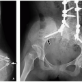
FIGURE 21.13 Polyostotic fibrous dysplasia in a 9-year-old boy with unilateral distribution in the appendicular skeleton and facial involvement. A: Lateral skull radiograph demonstrates facial deformity secondary to fibrous dysplasia with characteristic ground-glass expansile lesions of the base of the skull and frontal bone, maxilla, and the anterior mandible (arrows). B: Frontal radiograph of the pelvis demonstrates coxa vara deformity with expansile mixed-sclerotic fibrous dysplasia lesion in the right femoral neck (arrow). A similar smaller fibrous dysplasia lesion is noted in the medial aspect of the left proximal femoral diaphysis (interrupted arrow). C: Frontal view of the right humerus demonstrates an expansile ground-glass lesion of the mid humeral diaphysis with subtle endosteal scalloping and cortical thinning (arrow).

NF1 or von Recklinghausen disease has an incidence of 1 of 2,500 to 3,000 individuals.73,74 The disease is caused by an autosomal dominant mutation in the neurofibromin gene at 17q11.2 (OMIM 162200). The clinical manifestations of NF1 are extensive and involve multiple organ systems. The most common features include café-au-lait spots, Lisch nodules in the eye, cutaneous and deep plexiform neurofibromas, macrocephaly, optic glioma, and other neoplasms (both benign
and malignant). Musculoskeletal manifestations of NF1 include skull abnormalities (sphenoid wing dysplasia, erosions, and enlargement of foramina), scoliosis and kyphosis, pseudarthrosis of long bones (tibia most common), osteoporosis, and short stature (Fig. 21.15).72


FIGURE 21.14 Neurofibromatosis type 1. Plexiform neurofibroma, diffusely expanding a peripheral nerve and its branches (upper panel), is seen. The cut surface (lower left) is tan white and firm, resembling fibrous tissue. In this example, the majority of the mass has the histologic appearance of neurofibroma (lower center), whereas areas of increased cell density and round “epithelioid” cells characterize a component of malignant peripheral nerve sheath tumor (lower right). (Hematoxylin and eosin, original magnification, 400×.)

Only gold members can continue reading. Log In or Register to continue

Stay updated, free articles. Join our Telegram channel

Oct 13, 2018 | Posted by in PEDIATRIC IMAGING | Comments Off on Normal Growth, Normal Development, and Congenital Disorders

Full access? Get Clinical Tree

Get Clinical Tree app for offline access