and Marguerite M. Caré1
(1)
Department of Radiology ML5031, Cincinnati Children’s Hospital, Cincinnati, Ohio, USA
Keywords
Child abuseMetaphyseal collarBone barkUlnas2.1 Metaphyseal Collar
Understanding the metaphyseal collar is one of the keys to diagnosing “normal” rather than “abuse” in evaluating the metaphyseal region of long bones in infants.
Professor Maurice Laval-Jeantet in Paris may well have been the first investigator to recognize the metaphyseal collar (Figs. 2.1, 2.2, 2.3, and 2.4) during his investigation of the diaphyses (shafts) of long bones of experimental newborn animals (personal communication).
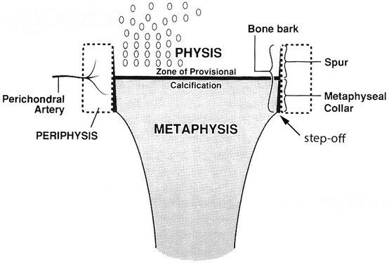
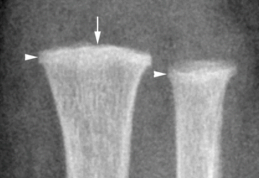


Fig. 2.1
Artist rendition of the region of the metaphyseal collar and the zone of provisional calcification. Note the step off between the straight metaphyseal collar (formed by the bone bark) and the curved diametaphysis beyond it. The spur is that part of the one-cell-layer-thick bone bark surrounding the most mature (cartilaginous) physis (Reprinted from Oestreich and Ahmad [4])
Fig. 2.2
The straight metaphyseal collar (arrowheads) of the distal radius and ulna of an infant. The arrow indicates the site of the calcified cartilaginous zone of provisional calcification
Fig. 2.3
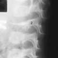
The straight metaphyseal collar (arrow) at the distal tibia of a 6-month-old girl
Fig. 2.4
Ultrasound imaging also clearly demonstrates bone bark at the metaphyseal collar (arrowhead) and the zone of provisional calcification (arrow) in a 7-month-old infant
Most of the membranous bone of tubular bones, and their flat bone equivalents, is produced from periosteum laying down cortical bone. (Reminder: epiphyses and their equivalents including tarsal and carpal bones do not have a periosteum, and so do not create membranous bone.) However, a special type of membranous bone formation occurs at the ends of the shafts of tubular bones – there a one-cell-layer-thick membranous bone, called bone bark, is formed. In general, part of the bark surrounds the physis (growth plate) immediately proximal to the metaphysis and part of the bark surrounds the most recently created metaphysis (Figs. 2.5 and 2.6). The mineralized bone bark is visible as a straight line at the edge of the most mature physis at the wrist in the first year of life in about 10 % of studies (Fig. 2.7). The result of the bone bark is a straight margin of that most recent metaphysis, which surrounds it circumferentially. A collar is thus formed at the extremity of the shaft. It is generally 1–3 mm in longitudinal width. Although the collar is present in the metaphyseal region of all long tubular bones, it is best visualized at the distal radius and ulna, and also seen well at the distal tibia and fibula. The anatomy of the proximal femur makes the collar difficult to appreciate there, however. For any site, the collar is better seen on an image centered at the site rather than on a larger view centered at another portion of the bone (see Fig. 1.3). This collar is present at birth and remains present until later childhood [1]. At the junction of the collar with the shaft (periosteal) membranous bone is found, therefore, an abrupt contour change between the straight margin and the curved margin which slowly narrows the shaft on the way to the middiaphysis. That contour change (Kleinman calls it a stepoff ) is a normal finding not to be mistaken for an abuse buckle fracture. Note, the classic metaphyseal corner fracture occurs in a similar region of the bone, emphasizing the importance of recognizing the normal and not misdiagnosing it.


Fig. 2.5
Photomicrograph of an (animal) specimen shows the bone bark spur (A to B) and the bone bark metaphyseal collar (B to C) as well as the transverse zone of provisional calcification (arrowheads) (Modified from Brighton [5], with permission)
Fig. 2.6
Specimen radiograph of a newborn animal showing the bone bark in profile (arrow 1) and en face (arrow 2) and the zone of provisional calcification (arrowheads) (Courtesy of Dr. Elfi Divanli)
One of the more difficult tasks in the evaluation for child abuse is recognizing the metaphyseal corner irregularities that are normal, due to a prominence of bone bark, often slightly separated from the adjacent bony contour. These irregularities have a pattern not much different from metaphyseal corner fractures which comprise classic metaphyseal lesions. I am afraid that I cannot state any distinguishing features to make a certain pronouncement. Sometimes, the lack of change of a follow up radiograph some 11–14 days later is reassuring for normality; sometimes the follow up will show change and even periosteal reaction that will affirm a traumatic etiology. Ultrasound images tend to show the same pattern as radiographs, but might show periosteal elevation as a sign of fracture. I here show three examples that by my experience are examples of the normal bone bark related irregularity (Fig. 2.8a–c). Next, I show a 3-day-old’s radius which has a metaphyseal prominence to its lateral contour (Fig. 2.8d) which is more like a classic metaphyseal lesion (presumably from birth trauma) than the bone bark just discussed. Indeed, on follow up 13 days later, periosteal reaction has appeared, confirming it was a corner fracture (Fig. 2.8e).