Normal Vessel Anatomy


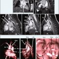
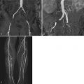
  • Optimal visualization of normal vascular anatomy with magnetic resonance angiography can be achieved with gadolinium-enhanced three-dimensional (3D) techniques, phased array surface coils, and parallel imaging.


  • Three-dimensional reconstruction techniques are typically used and consist of multiplanar reformatted images and full or subvolume maximum intensity projection images in various planes.


KEY POINTS


This chapter reviews the normal arterial and venous anatomy as well as some of the more common normal variants for the extracranial circulation in the form of an anatomic atlas. All images were obtained on state-of-the-art 1.5T and 3T whole-body scanners. Gadolinium-enhanced 3D techniques were used for all cases using phased array surface coils and parallel imaging. To optimally visualize the individual vascular territories, the most appropriate 3D reconstruction techniques were used. These consist of multiplanar reformatted images and full or subvolume maximum intensity projection images in various planes, in addition to source images.


Figure 2-1


Coronal maximum intensity projection (MIP) image of contrast-enhanced magnetic resonance angiogram at 3 Tesla (3T contrast-enhanced MRA) of the supraaortic arteries in a 78-year-old man. The three main supraaortic branches include the brachiocephalic, left common carotid, and left subclavian arteries, which supply the head and upper extremities.


Figure 2-2


Oblique sagittal MIP image of 3T contrast-enhanced MRA of the left carotid artery in a 78-year-old man. The internal carotid artery has a more posterior and lateral course as compared with the external carotid artery. There are no branches of the internal carotid artery below the skull base (cervical portion).


Figure 2-3


Coronal MIP image of 3T contrast-enhanced MRA of the vertebral arteries in a 78-year-old man. In the majority of patients, there is a dominant vertebral artery that is larger than its contralateral counterpart. In this case, the left vertebral artery is dominant.


Figure 2-4


Coronal MIP image of 1.5T contrast-enhanced MRA of the upper extremity arterial tree in a 74-year-old man. The right subclavian artery originates at the bifurcation of the brachiocephalic artery and right common carotid artery, extending to the point where it crosses the outer border of the first rib, where it becomes the axillary artery. The axillary artery continues to the lower border of the tendon of the teres major muscle, where it becomes the brachial artery.


Figure 2-5


Left anterior oblique MIP image of 1.5T contrast-enhanced MRA of the upper extremity arterial tree in a 74-year-old man. The ascending aorta is comprised of the aortic root (sinuses of Valsalva) and the tubular portion. The aortic arch gives rise to the supraaortic branches (see Fig. 2-1 ). The descending aorta begins in the region of the ligamentum arteriosum (remnant of the ductus arteriosus).


Figure 2-6


Coronal MIP image of 1.5T contrast-enhanced MRA of the pulmonary arteries in a 25-year-old woman. The main pulmonary artery bifurcates into left and right pulmonary arteries. The right pulmonary artery courses to the right, whereas the left pulmonary artery continues posteriorly.


Figure 2-7


Sagittal subvolume MIP image of 1.5T contrast-enhanced MRA of the right pulmonary artery (RPA) in a 25-year-old woman. The RPA gives rise to ascending and descending branches, with the ascending branch supplying the segmental arteries of the upper lobe. An accessory branch to the right upper lobe is present in approximately 90% of individuals. The descending branch gives rise to the segmental branches of the right middle and lower lobes.


Figure 2-8


Sagittal subvolume MIP image of 1.5T contrast-enhanced MRA of the left pulmonary artery in a 25-year-old woman. The left pulmonary artery (LPA) is a little shorter and smaller than the RPA. It runs horizontally in front of the descending aorta and left bronchus to the root of the left lung, where is divides into two branches: one for upper lobe (gives off apical, posterior, anterior descending, anterior descending, and lingular arteries) and one for lower lobe (gives off superior “apical,” medial basal, anterior basal, lateral basal, and posterior basal arteries).


Figure 2-9


Sagittal subvolume MIP image of 1.5T contrast-enhanced MRA of the thoracic aorta in a 29-year-old woman. The bronchial arteries are quite variable, and can arise directly from the aorta or a common trunk with posterior intercostal arteries. The most superior posterior intercostal arteries arise either from the costocervical trunk (branch of the subclavian artery) or common trunks arising from the proximal descending aorta. The more inferior posterior intercostal arteries arise in left and right pairs from the descending aorta at each level.


Figure 2-10


Coronal subvolume MIP image of 1.5T contrast-enhanced MRA of descending thoracic aorta in a 29-year-old woman. Three types of intercostal arteries are present: superior intercostal arteries (from the costocervical trunk and supplying the first and second posterior intercostal spaces), anterior intercostal arteries (six pairs arising from the internal thoracic artery and supplying the upper six intercostal spaces), and posterior intercostal arteries (nine pairs arising from the thoracic aorta and supplying the lower nine intercostal spaces). The intercostal arteries originate separately, one on each side, from the descending thoracic aorta (80% of individuals), or from a common trunk. A unilateral trunk is present in 10% to 15% of individuals.


Figure 2-11


Coronal MIP image of 3T contrast-enhanced MRA of abdominal aorta in a 24-year-old woman. The abdominal aorta begins at the aortic hiatus of the diaphragm and ends normally at the level of the body of the fourth lumbar vertebra by dividing into the two common iliac arteries. (Occasionally, the aortic bifurcation is higher or lower.) Its branches may be divided into four sets: dorsal (supplying the body wall), ventral and lateral (supplying the viscera), and terminal (supplying pelvis and lower limbs).


Figure 2-12


Right anterior oblique MIP image of 3T contrast-enhanced MRA of abdominal aorta in a 34-year-old woman. The ventral branches of the abdominal aorta include celiac, superior mesenteric, and inferior mesenteric arteries. The dorsal branches include lumbar and median sacral arteries. The common iliac arteries are the terminal branches of the abdominal aorta. (Absent or very short common iliac arteries are rare.)


Figure 2-13


Sagittal subvolume MIP image of 3T contrast-enhanced MRA of abdominal aorta in a 62-year-old woman. Lumbar arteries (usually four on each side) arise from the back of the aorta opposite the bodies of the upper four lumbar vertebrae. A fifth pair, smaller in size, occasionally arises from the median sacral artery, but the lumbar branches of iliolumbar arteries usually take their place.


Figure 2-14


Coronal subvolume MIP image of 3T contrast-enhanced MRA of celiac and mesenteric arteries in a 47-year-old woman. The celiac trunk arises at the T12-L1 level and is usually directed inferiorly but can be horizontal or craniad. Classically, (in 65% to 75% of individuals) it divides into left gastric (usually the first branch), common hepatic, and splenic arteries. The superior mesenteric artery (SMA) origin is located around L1, slightly below the celiac artery (range, 2 mm to 2 cm). It supplies the whole of the small intestine (except the superior part of the duodenum), cecum, the ascending colon and most of the transverse colon. The SMA gives off inferior pancreaticoduodenal, ileocolic, right colic, middle colic, jejunal, and ileal branches.

Only gold members can continue reading. Log In or Register to continue

Stay updated, free articles. Join our Telegram channel

Feb 1, 2019 | Posted by in MAGNETIC RESONANCE IMAGING | Comments Off on Normal Vessel Anatomy

Full access? Get Clinical Tree

Get Clinical Tree app for offline access