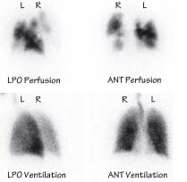
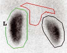
The perfusion pattern matches the ventilation pattern. The probability of a significant perfusion defect is therefore low. LPO left posterior oblique, ANT anterior
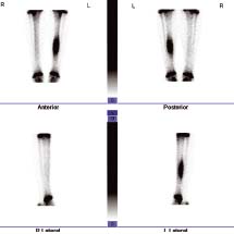
50.2 V/Q scan: high probability of pulmonary embolus
This patient presented with sudden breathlessness. Ventilation is normal but perfusion is patchy, indicating a high probability of pulmonary embolus. Anticoagulation therapy was initiated
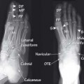
50.3 Bone scan
(Same patient as fig 50.4.) This child presented with left tibial pain, worse at night, and relieved with simple analgesia. These are typical clinical features of an osteoid osteoma. The plain XR showed cortical thickening only. This bone scan shows high uptake of radionuclide material at the site of pain, which indicates an active inflammatory lesion
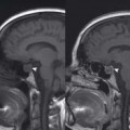
50.4 CT v PET-CT
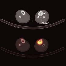
(Same patient as fig 50.3.) The CT scan (top) shows the small lucent ‘nidus’ of an osteoid osteoma (a benign lesion) with surrounding reactive bone formation (arrowheads). The bottom image is a PET/CT scan clearly indicating the location of the active lesion
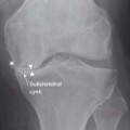
50.5 Bone scan
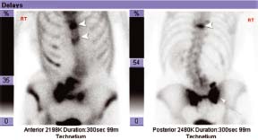
This patient with osteoporosis presented with increasing lower back pain. Plain XRs showed vertebral insufficiency fractures which are also seen on these images (arrowheads) but did not reveal the bilateral sacral insufficiency fractures as is evident by uptake in the shape of an H on this scan (Honda sign)
50.6 DMSA: posterior view
This is a DMSA study in a patient with renal artery stenosis. It was performed to determine comparative functional uptake of radionuclide contrast material. In this case the right kidney showed a relative uptake of 42%
Ventilation/Perfusion (V / Q) scanning
V/Q scanning is used to assess the flow of air and blood in the lungs. The ventilation phase involves the patient inspiring xenon or technetium-99m labelled diethylenetriamine pentaacetic acid (99m Tc DTPA) and the perfusion phase involves the injection of technetium-99m labelled macroaggregated albumin (99m
Related posts:


Stay updated, free articles. Join our Telegram channel

Full access? Get Clinical Tree


