Hernia most commonly contains loop of Ileum, although can rarely involve other pelvic viscera (i.e., bladder)
Most often trapped between obturator externus and pectineus muscles
May also be located between superior and middle fasciculi of obturator externus or between internal and external obturator muscles
• Hernia sac exits pelvis near obturator vessels and nerve
• Right side more common
TOP DIFFERENTIAL DIAGNOSES
• Inguinal hernia
• Sciatic hernia
• Perineal hernia
• Femoral hernia
PATHOLOGY
• Defect in pelvic floor or laxity of pelvic muscles and fascia
• Made worse by any chronic increase in abdominal pressure (COPD, constipation, pregnancy, etc.)
• More common in thin or emaciated patients, as preperitoneal fat usually supports obturator canal
CLINICAL ISSUES
• Accounts for < 1% of all hernias
• > 90% occur in elderly women (mean age 82)
Less common complication of pelvic floor laxity
• Acute or recurrent small bowel obstruction, partial > complete
80% of patients present with symptoms of bowel obstruction
Majority require resection of strangulated small bowel
• Rare occurrence and nonspecific signs often lead to late diagnosis
Correct clinical diagnosis in only 10–30% of cases
Diagnosis best made by CT/MR rather than clinical exam
(Left) Graphic shows a bowel obstruction caused by an obturator hernia. Strangulated bowel lies deep to the pectineus muscle and superficial to the obturator externus muscle .
(Right) Axial CECT in a 73-year-old woman shows a protrusion of portions of the bladder into bilateral obturator hernias . Obturator hernias most commonly contain herniated ileum, but other pelvic viscera can also herniate, as in this case.
(Left) Axial CECT in a 90-year-old woman with bowel obstruction shows dilated proximal small bowel loops and collapsed distal bowel .
(Right) Axial CECT in the same patient shows the herniated and strangulated segment of the ileum trapped between the obturator externus and the pectineus muscles. These are the classic imaging findings of an obturator hernia.
TERMINOLOGY
Abbreviations
• Obturator hernia (OH)
Definitions
• Pelvic hernia protruding through obturator foramen
IMAGING
General Features
• Best diagnostic clue
CT evidence of herniated bowel lying between pectineus and obturator muscles in an elderly woman
Radiographic Findings
• Abdominal radiographs or barium studies
Small bowel obstruction with a fixed loop containing gas or contrast medium in obturator region
Only gold members can continue reading. Log In or Register to continue

 May also be located between superior and middle fasciculi of obturator externus or between internal and external obturator muscles


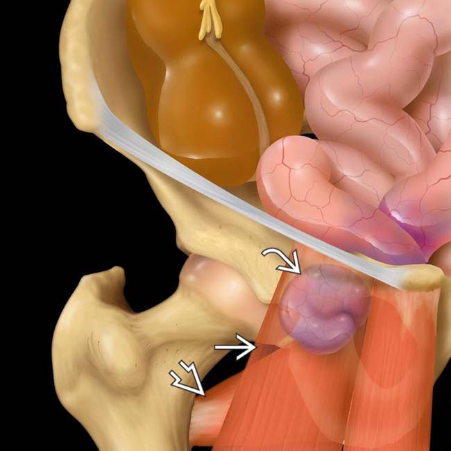
 lies deep to the pectineus muscle
  and superficial to the obturator externus muscle
  .
.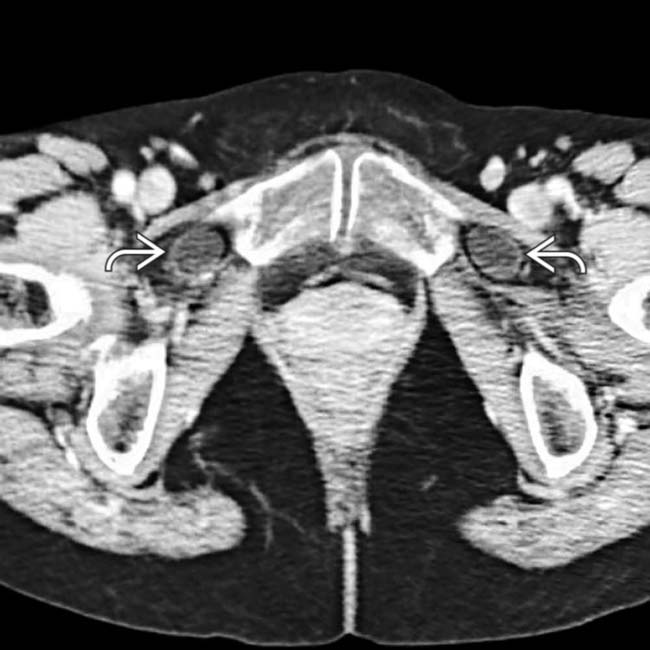
 . Obturator hernias most commonly contain herniated ileum, but other pelvic viscera can also herniate, as in this case.
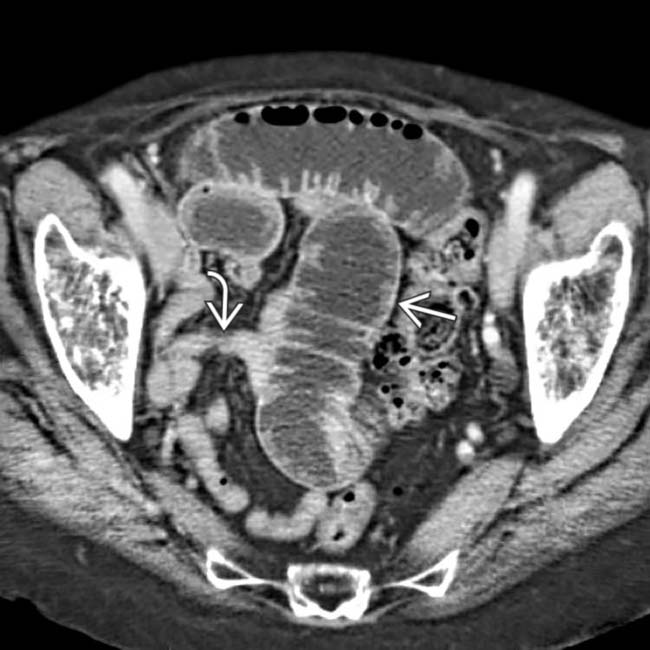
 and collapsed distal bowel
  .
.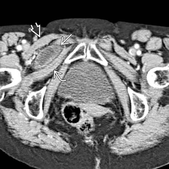
 trapped between the obturator externus
  and the pectineus
  muscles. These are the classic imaging findings of an obturator hernia.


