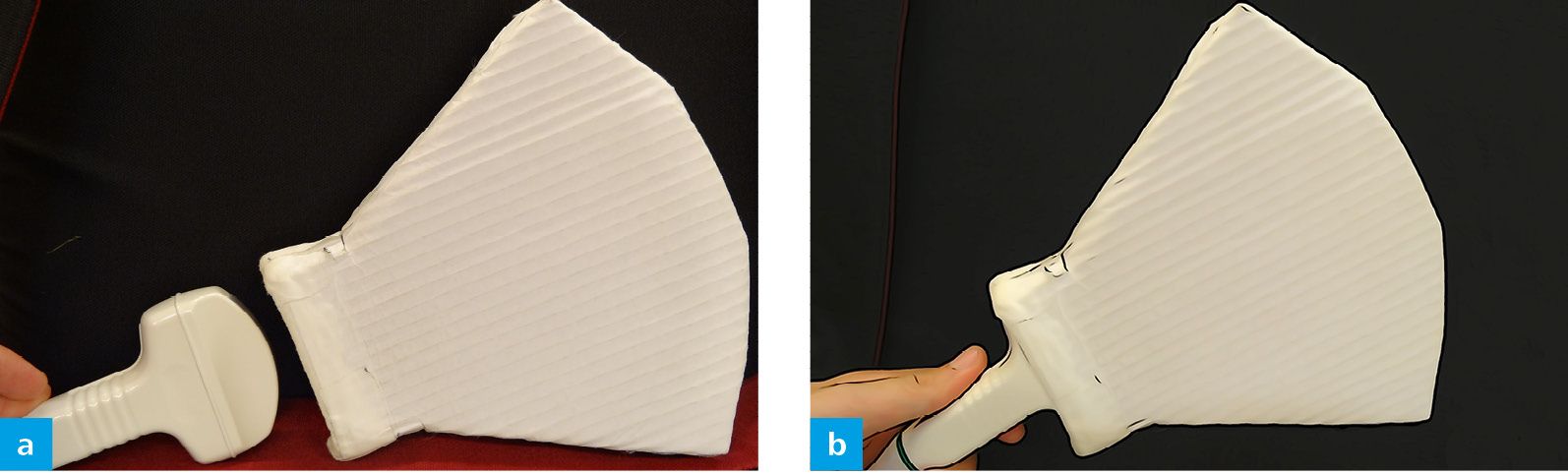
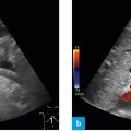
Fig. 1: Convex probe 2 – 6 MHz (a), linear probe 6 – 15 MHz (b), and high-resolution linear “hockey stick” to – 18 MHz – for special applications (c).
In ultrasound we use various types of probes, which are named according to the propagation of the ultrasound waves from the probe to the periphery. A convex probe is used for ultrasound investigation of the abdomen (Fig. 2a). It emits ultrasound waves in a fan-shaped manner at a frequency of 2 – 6 MHz from the surface of the probe. A low frequency of this nature is required for the ultrasound wave to reach a depth of 10 – 15 cm and produce an adequate picture by reflection and registration at the probe.
Related posts:

Stay updated, free articles. Join our Telegram channel

Full access? Get Clinical Tree


