= from nasion to anterior angle of bregma
[bregma, Greek = top of head]
Closure: by 3 months – 6 years of age; in up to 10% open until adulthood
Sutura frontalis persistens = metopism
= no closure of incomplete / complete metopic suture
DDx: anterior vertical fracture
B. SAGITTAL SUTURE
[sagitta, Latin = arrow]
= fibrous connective tissue joint between two parietal bones
Average width: 5.0 ± 0.2 mm (at birth), 2.4 ± 0.1 mm (1 month of age); narrowing further over time
Closure: 21–30 years of age; fusing anteriorly beginning at intersection with lambdoid suture
C. CORONAL SUTURE
= separates frontal from parietal bones
Average width: 2.5 ± 0.1 mm (at birth), 1.3 ± 0.1 mm (1 month of age)
Closure: 24 years of age
D. SQUAMOSAL SUTURE
(a) temporosquamosal suture
= connects temporal bone squama with lower border of parietal bone; arches posteriorly from pterion (= contact point between frontal, parietal, temporal, sphenoid)
[pteron, Greek = wing]
√ often visualized at two points at CT with lambdoid suture acting as a useful posterior reference point
= continuous posteriorly with parietomastoid suture uniting mastoid process of temporal bone with region of mastoid angle of parietal bone
(b) sphenosquamosal suture
= courses inferiorly from pterion separating sphenoid bone from squama of temporal bone
N.B.: often mistaken for skull base fracture
E. LAMBDOID SUTURE
[upper case Greek letter lambda = L]
= connects parietal with occipital bone
Closure: 26 years of age
N.B.: the most common site of wormian bones
F. OCCIPITOMASTOID SUTURE
= inferior continuation of lambdoid suture at the point where lambdoid suture intersects with temporosquamosal suture
G. PARIETOMASTOID SUTURE
= links temporosquamosal and lambdoid sutures
√ often not seen on axial CT images
H. OCCIPITOMASTOID SUTURE
= between occipital bone + mastoid process of temporal bone as a continuation of the lambdoid suture toward skull base
N.B.: not infrequently mistaken for a skull base fracture
I. SPHENOFRONTAL SUTURE
= transverse suture between anterior margin of lesser sphenoid wing + posterior margin of horizontal orbital plate
√ lesser sphenoid wing (posterior to suture) is a useful landmark for suture localization
J. ACCESSORY PARIETAL SUTURE (RARE)
= the most common of all usually bilateral and symmetric accessory sutures
Location: parietal and occipital bone → multiple ossification centers
K MENDOSAL / ACCESSORY OCCIPITAL SUTURE
Frequency: 3% in an Indian subcontinent population
Closure: in utero / first few days of life; may persist up to 6 years of age
Os incae = large single centrally located intrasutural bone at junction of lambdoid and sagittal sutures; often forms in a persistent mendosal suture
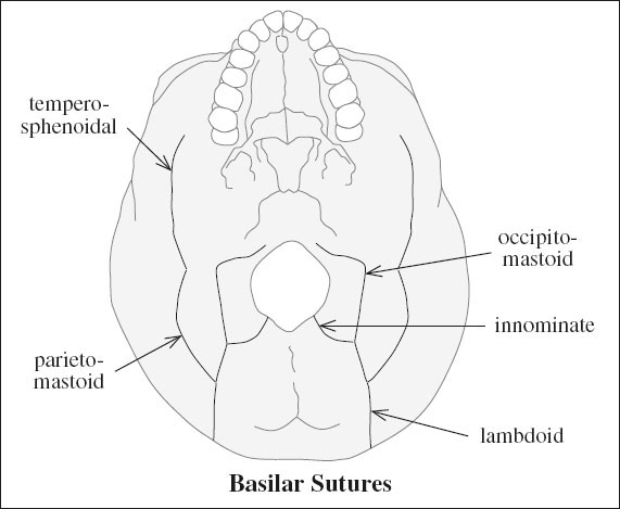
K SKULL BASE SUTURES
Ossification: 50% (84%) of anterior base by 6 (24) months
(a) innominate / intraoccipital
Closure: 4 years of age
(b) lambdoid
(c) occipitomastoid
(d) parietomastoid
(e) temporosphenoidal
Symmetry and knowledge of the anatomic appearances of basal sutures are important for avoiding misdiagnosis.
A persistent hypoattenuating area of any length extending from foramen magnum beyond 4 years of age indicates a fracture.
FORAMINA OF BASE OF SKULL
on inner aspect of middle cranial fossa 3 foramina are oriented along an oblique line in the greater sphenoidal wing from anteromedial behind the superior orbital fissure to posterolateral
mnemonic: “rotos”
foramen rotundum
foramen ovale
foramen spinosum
Foramen Rotundum
= canal within greater sphenoid wing connecting middle cranial fossa + pterygopalatine fossa
| Location: | inferior and lateral to superior orbital fissure | |
| Course: | extends obliquely forward + slightly inferiorly in a sagittal direction parallel to superior orbital fissure | |
| Contents: | (a) nerves: | V2 (maxillary nerve) |
| (b) vessels: | (1) artery of foramen rotundum | |
| (2) emissary vv. | ||
√ best visualized by coronal CT
Foramen Ovale
= canal connecting middle cranial fossa + infratemporal fossa
Location: medial aspect of sphenoid body, situated posterolateral to foramen rotundum (endocranial aspect) + at base of lateral pterygoid plate (exocranial aspect)
| Contents: | (a) nerves: | (1) V3 (mandibular nerve) |
| (2) lesser petrosal nerve (occasionally) | ||
| (b) vessels: | (1) accessory meningeal artery | |
| (2) emissary veins |
Foramen Spinosum
Location: on greater sphenoid wing posterolateral to foramen ovale (endocranial aspect) + lateral to eustachian tube (exocranial aspect)
| Contents: | (a) nerves: | (1) recurrent meningeal branch of mandibular nerve |
| (2) lesser superficial petrosal nerve | ||
| (b) vessels: | (1) middle meningeal artery | |
| (2) middle meningeal vein |
Foramen Lacerum
covered (occasionally) by fibrocartilage, carotid artery rests on endocranial aspect of fibrocartilage
Location: at base of medial pterygoid plate
Contents: (inconstant)
(a) nerve: pterygoid canal n. (actually pierces cartilage)
(b) vessel: meningeal branch of ascending pharyngeal a.
Foramen Magnum
| basion | = | anterior lip of foramen |
| opisthion | = | posterior lip of foramen |
| Contents: | (a) nerves: | (1) medulla oblongata |
| (2) CN XI (spinal accessory nerve) | ||
| (b) vessels: | (1) vertebral artery | |
| (2) anterior spinal artery | ||
| (3) posterior spinal artery |
Pterygoid Canal
= VIDIAN CANAL
= within sphenoid body connecting pterygopalatine fossa anteriorly to foramen lacerum posteriorly
Location: at base of pterygoid plate below foramen rotundum
Contents: (a) nerves: vidian nerve = nerve of pterygoid canal = continuation of greater superficial petrosal nerve (from cranial nerve VII) after its union with deep petrosal nerve
(b) vessel: vidian artery = artery of pterygoid canal = branch of terminal portion of internal maxillary a. arising in pterygopalatine fossa → passing through foramen lacerum posterior to vidian n.
Hypoglossal Canal
= ANTERIOR CONDYLAR CANAL
Location: in posterior cranial fossa anteriorly above condyle starting above anterolateral part of foramen magnum, continuing in an anterolateral direction + exiting medial to jugular foramen
| Contents: | (a) nerves: | cranial nerve XII (hypoglossal n. |
| (b) vessels: | (1) pharyngeal artery | |
| (2) branches of meningeal artery |
Jugular Foramen
Location: at posterior end of petrooccipital suture directly posterior to carotid orifice
(a) anterior part:
(1) inferior petrosal sinus
(2) meningeal branches of pharyngeal artery + occipital a.
(b) intermediate part:
(1) cranial nerve IX (glossopharyngeal nerve)
(2) cranial nerve X (vagus nerve)
(3) cranial nerve XI (spinal accessory nerve)
(c) posterior part: internal jugular vein
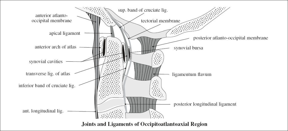
CRANIOVERTEBRAL JUNCTION (CVJ)
CRANIOCERVICAL JUNCTION: C1 (atlas) + C2 (axis) + occiput
Variants of CVJ: precondylar tubercles, third occipital condyle, ossification of ligament of odontoid process
Craniometry:
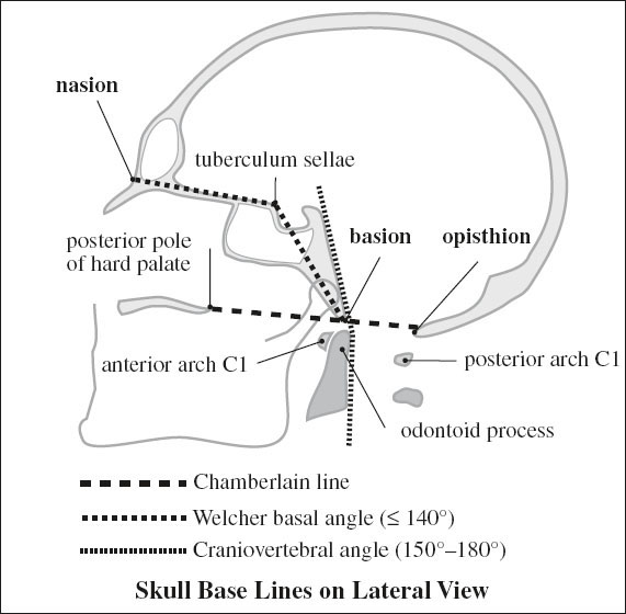
› LATERAL VIEW
1. Chamberlain line
= line between posterior edge of hard palate + posterior margin of foramen magnum (= opisthion)
√ tip of odontoid process usually lies below / tangent to Chamberlain line by > 3 mm
√ tip of odontoid process may lie up to 1 ± 6.6 mm above the Chamberlain line
2. McGregor line
= line between posterior edge of hard palate + most caudal portion of occipital squamosal surface
◊ Substitute to Chamberlain line if opisthion not visible
√ tip of odontoid < 4.5–5.0 mm above this line
3. Wackenheim clivus baseline
= BASILAR LINE = CLIVAL LINE = line along clivus
√ usually falls tangent to posterior aspect of tip of odontoid process
4. Craniovertebral angle = clivus-canal angle
= angle formed by line along posterior surface of axis body and odontoid process + basilar line
√ ranges from 150° in flexion to 180° in extension
√ ventral spinal cord compression may occur at < 150°
5. Welcher basal angle
= intersection of nasion-tuberculum line and of tuberculum-basion line (along clivus)
√ angle averages 132° (should be < 140–145°)
6. McRae line
= line between anterior lip (= basion) to posterior lip (= opisthion) of foramen magnum
√ tip of odontoid below this line = NO basilar invagination; if poorly seen → Chamberlain line
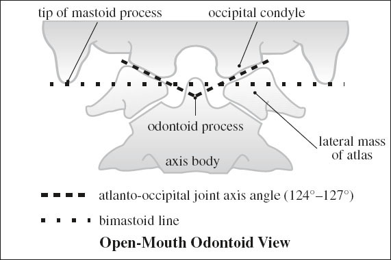
› ANTEROPOSTERIOR VIEW (= “open-mouth” / odontoid view)
7. Atlanto-occipital joint axis angle
= formed by lines drawn parallel to atlantooccipital joints
√ lines intersect at center of odontoid process
√ average angle of 125° (range, 124° to 127°)
Related posts:


Stay updated, free articles. Join our Telegram channel

Full access? Get Clinical Tree


