, Valery Kornienko2 and Igor Pronin2
(1)
N.N. Blockhin Russian Cancer Research Center, Moscow, Russia
(2)
N.N. Burdenko National Scientific and Practical Center for Neurosurgery, Moscow, Russia
Oligodendroglioma (ODG) is a relatively rare glioma, also quite a demarcated tumor. Moreover, “pure” ODGs are even more rare; mostly mixed gliomas are observed (oligoastrocytomas). The tumor develops from a specific type of glial cells—oligodendrocytes—and accounts for 2–10% of all primary intracranial tumors and 5–25% of gliomas. Most of ODGs are slow-growing, benign, non-encapsulated infiltrative tumors of the white matter, which are usually located paraventricularly. 85% of tumors are supratentorial, affecting mostly the frontal lobe. The tumor is characterized by the formation of foci of cystic degeneration and calcifications. Malignization of ODGs manifests by cellular atypia, occurrence of mitoses in the tumor, and proliferation of vascular endothelium (Fig. 29.1).
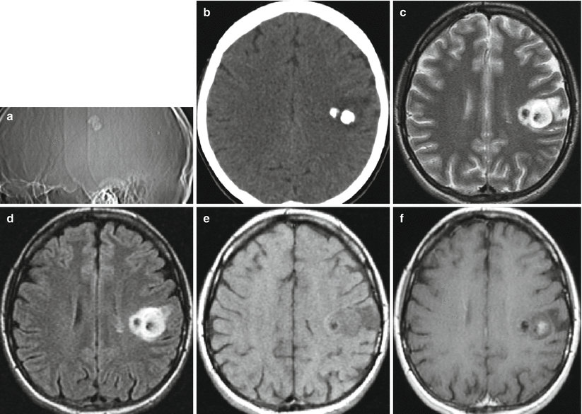
Fig. 29.1
Oligodendroglioma. In the left postfrontal area, on the lateral radiograph (a) and CT in the axial projection (b), two adjacent calcifications are visualized. On CT, there is additionally an area with decreased brain tissue density, surrounding calcifications. In a series of T2-weighted (c), T2-FLAIR (d), T1-weighted MRI before (e) and after intravenous contrast enhancement (f), a space-occupying lesion is determined, having mostly an increased MR signal (T2-weighted, T2-FLAIR MRI) with portions of the reduced signal (calcifications) and a local area of abnormal CA accumulation
Oligodendroglioma is a most common intracranial tumor that calcifies, which is clearly seen on CT. The neoplasm has a mixed density. 75% of ODGs do not accumulate contrast agent. MRI identifies a tumor with mixed hypo- and isointensity in T1-weighted images and hyperintense foci in T2-weighted images (Fig. 29.2).
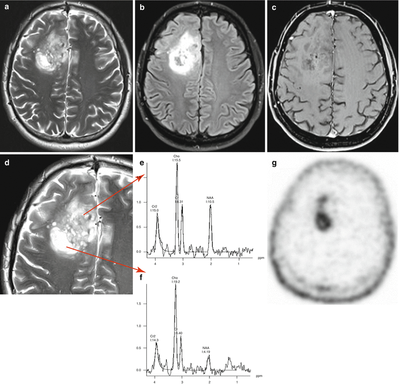
Fig. 29.2


Oligodendroglioma. In the right frontal region in T2-weighted (a, d) and T2-FLAIR MRI (b) images, there is a large space-occupying lesion with a heterogeneously high signal. After CA administration, on T1-weighted (c), there are no signs of its accumulation in the tumor. On the spectra from the anterior (e) and posterior poles (f) of the tumor, benign and malignant characteristics are observed, respectively (appearance of the Lip-Lac complex peak and a pronounced increase in the Cho peak). On PET with 18F-tyrosine, tumor fragments are well differentiated with different RP accumulation, corresponding to various malignancy grades (g)
Related posts:


Stay updated, free articles. Join our Telegram channel
Full access? Get Clinical Tree
