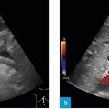
Fig. 1: Holding the probe for a longitudinal section: the red line highlights the marker.
When obtaining a standardized longitudinal section, the marker on the probe (red line) always points in cranial direction.
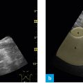
The probe is held perpendicular to the skin and a sagittal section is obtained.
Thus, the left side of the image is oriented cranially and the right side is oriented caudally. Close to the probe is the ventral aspect, which is identical with the upper part of the image. The portion farther away from the probe is the dorsal aspect; it is identical with the lower part of the image. The orientation of the longitudinal section is always the same when the marker on the probe points strictly in cranial direction.
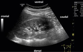
Fig. 2: Orientation of the sides of the image on the monitor in the longitudinal section: B-mode image
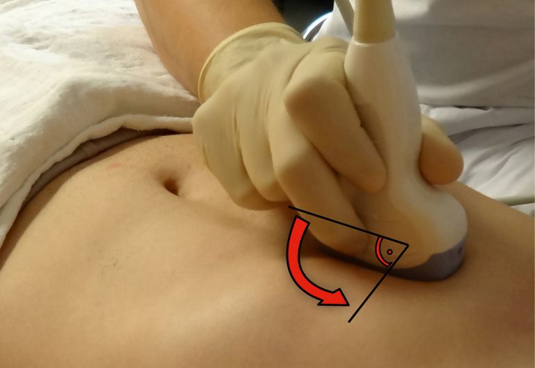
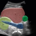
Fig. 3: Rotating the probe from the longitudinal section to the cross-section.
Related posts:


Stay updated, free articles. Join our Telegram channel

Full access? Get Clinical Tree


