Opening Round

Opening Round


Answers


Case 1


Lung Cancer


Comment


A solitary pulmonary nodule is defined as a well-circumscribed round or oval lesion measuring less than 3 cm in diameter. There are only two specific and reliable signs of benignancy on chest radiographs: (1) identification of a benign pattern of calcification or (2) demonstration of absolute absence of growth over a 2-year period. For cases that do not meet one of these criteria, thin-section CT is generally recommended for further evaluation. In comparison with radiography, CT allows a more accurate assessment of the margins of a nodule; moreover, CT is more sensitive for identifying the presence and distribution of calcium and fat within a nodule.


The nodule in this case has spiculated margins, a finding that is highly suspicious for malignancy. Depending on local practice patterns and clinical circumstances, a preoperative biopsy may be requested. The peripheral location of this nodule makes it best suited for a transthoracic needle biopsy.


The most common cell type of lung cancer is adenocarcinoma. It most often presents as a solitary, peripheral nodule with spiculated margins. Based on size criteria outlined in recent revisions to the TNM staging system for lung cancer, this 4.0-cm mass is a T2a lesion. The revisions establish the following size cut-off points for T designations: T1a: tumor ≤2 cm in greatest dimension; T1b: tumor >2 cm but ≤3 cm in greatest dimension; T2a: tumor >3 cm but ≤5 cm in greatest dimension; T2b: tumor >5 cm but ≤7 cm in greatest dimension; T3: tumor >7 cm (see Thoracic Radiology: THE REQUISITES, Table 11-1).


Notes


Answers


Case 2


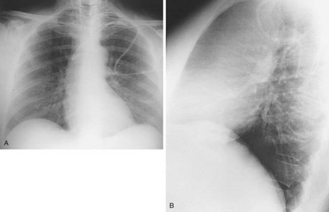
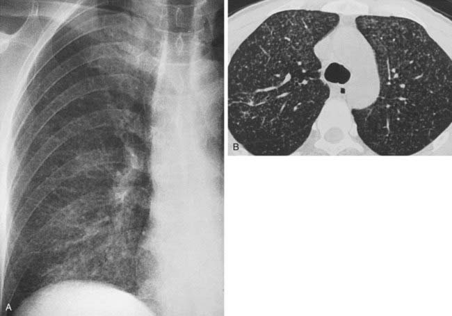
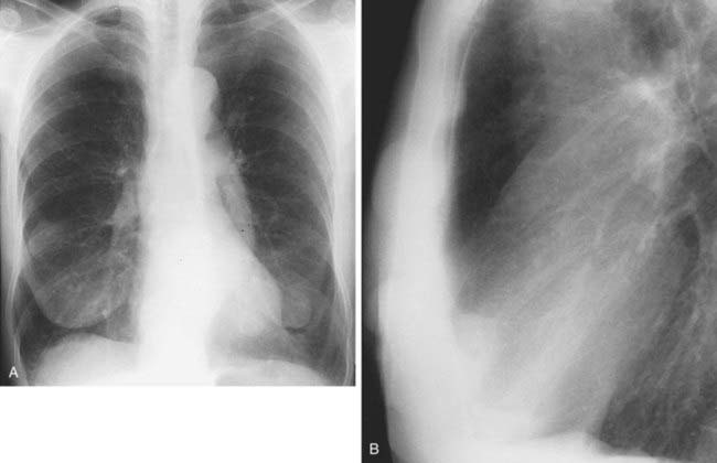
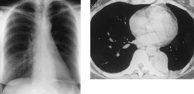
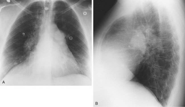
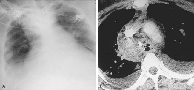
Spontaneous Pneumothorax Secondary to Ruptured Bleb


Comment


Pneumothorax is defined as the presence of air or gas within the pleural space. Although there are a wide variety of causes, spontaneous pneumothorax is the most common etiology. Affected patients are usually in the third or fourth decade of life.


Spontaneous pneumothoraces are almost always secondary to rupture of an apical bleb, which represents a gas pocket within the elastic fibers of the visceral pleura. Note the presence of a small bleb along the visceral pleural margin in this patient, best demonstrated on the coned-down image of the left upper lobe (arrow, second figure). Such blebs have been reported to be detectable on chest radiographs in approximately 15% of cases of spontaneous pneumothorax. However, blebs are rarely evident on chest radiographs following resolution of the pneumothorax. CT is much more sensitive than radiography for detecting blebs and has been shown to detect blebs in approximately 80% of patients following resolution of spontaneous pneumothoraces. The size and number of apical blebs detected on CT have been shown to correlate with the risk of recurrent pneumothoraces and the need for surgical intervention.


Tension pneumothorax is a life-threatening condition. Affected patients present with clinical signs of tachypnea, tachycardia, cyanosis, sweating, and hypotension. Radiographic findings may include contralateral mediastinal shift, diaphragmatic depression, rib cage expansion, and flattening of the contours of the right heart border and/or venae cavae.


Notes


Answers


Case 4


Calcified Pleural Plaques From Prior Asbestos Exposure


Answers


Case 5


Vertebral Body Fracture With Paraspinal Hematoma


Answers


Case 9


Complete Left Lower Lobe Atelectasis


Answers


Case 10


Primary Pulmonary Artery Hypertension


Answers


Case 11


Mediastinal Hematoma Secondary to Vascular Perforation by a Central Venous Catheter


Answers


Case 12


Sarcoidosis


Feb 28, 2016 | Posted by in RESPIRATORY IMAGING | Comments Off on Opening Round

Full access? Get Clinical Tree

Get Clinical Tree app for offline access