, Valery Kornienko2 and Igor Pronin2
(1)
N.N. Blockhin Russian Cancer Research Center, Moscow, Russia
(2)
N.N. Burdenko National Scientific and Practical Center for Neurosurgery, Moscow, Russia
Orbital sarcoidosis is a chronic multisystem granulomatous disease involving all organ systems. In case of systemic involvement, clinical manifestations of sarcoidosis occur in the CNS in 10% of cases (Mafee and Dorodi 1999). Typical features of the lacrimal gland involvement include an increase in its size, painless at palpation, and dry eyes. In case of involvement of the anterior chamber of the eye, symptoms include soreness, conjunctivitis, and uveitis; and involvement of the posterior chamber results to perivasculitis, infiltrates in the vitreous body and retinal changes, eyeball movement limitation, and diplopia. The optic nerve involvement manifests by perineuritis, papillitis, and loss of vision (Kellinghaus et al. 2004). Sarcoidosis more often occurs in adults aged from 30 to 50 years old, predominantly in women. It is ten times more frequently diagnosed among aborigines of the African deserts. Also, the disease is common in Puerto Ricans, Irish, and Scandinavians (Brovkina 2008).
An isodense increase in the nerve diameter is observed on CT before administration of the contrast material. After contrast enhancement, abnormal accumulation of the contrast agent by the optic nerve and other intraorbital structures is identified. In addition, IV contrast enhancement shows an increase of the lacrimal gland and diffuse irregular thickening of the extraocular muscles.
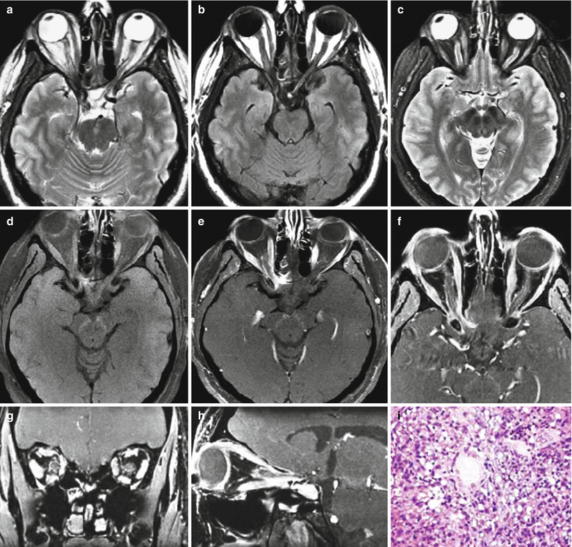
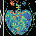
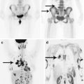
T1-weighted MRI sequences reveal an isointense enlargement of intraorbital structures. On T2-weighted images, hypointense (relative to normal muscle) or slightly hyperintense abnormal intraorbital masses can be visualized. T2 -FLAIR sequence can demonstrate sarcoidal parenchymal lesions in the brain. The optic nerve affected by orbital sarcoidosis and intraorbital granulomatous masses intensely accumulates the contrast agent. Contrast enhancement of the lacrimal gland and orbital muscles is also more pronounced than normal. In case of the CNS involvement, MRI shows contrast-enhanced intracranial cisternal and parenchymal lesions. Optimal scanning: study with contrast enhancement and suppression of fat MR signal. Not only the orbit should be examined but also the whole brain in order to identify other foci of sarcoidosis (Figs. 38.1 and 38.2).
