, Valery Kornienko2 and Igor Pronin2
(1)
N.N. Blockhin Russian Cancer Research Center, Moscow, Russia
(2)
N.N. Burdenko National Scientific and Practical Center for Neurosurgery, Moscow, Russia
According to publications, more than 100 different types of lesions can develop in the orbit (Rootman 1988; Baert 2006). The main role in the diagnosis of orbital lesions plays the analysis of history and clinical symptoms, and only a small proportion of cases requires neuroradiological studies to clarify the diagnosis. CT examinations of the orbit use mainly thin-slice helical scanning with reformations in the plane of interest. The use of CT perfusion imaging became a new approach in the diagnosis of orbital lesions, which opened a new era in the evaluation of hemodynamics of orbital tumors. MRI studies of the orbit are distinguished by the mandatory use of fat suppression techniques, both before and after intravenous contrast enhancement. Preference is given to T2- and T1-weighted sequences with thin 3-millimeter slices, directed along the course of the optic nerves in the axial, sagittal, and oblique views. Frontal projections should be oriented perpendicularly to the optic nerve; therefore, they are performed at least twice. Metastasizing of malignant tumors to the orbit is an uncommon event; however, attention should be paid to this area in melanoma and breast cancer.
37.1 Neurofibromas (NF)
Neurofibromas (NF) can be solitary, plexiform, and diffuse. Solitary NF usually develops in adults, while plexiform NF (typical for type I neurofibromatosis) develops within the first 5 years of life (Brovkina 1993). Plexiform (or reticular) neurofibromas are the most frequent in the orbit.
On CT, a single NF looks like a rounded, oblong, or oval encapsulated formation with the density close to that of the brain tissue, but this pattern, in general, is nonspecific; it has a medium degree of contrast enhancement.
On MR, NF has a homogeneous structure with irregular outlines: the tumor is usually represented by an infiltrative mass with irregular contours, which sometimes infiltrate the orbital adipose tissue. It is homogeneous and iso- or hypointense relative to the muscle tissue in T1-weighted images. On T2-weighted images, it can be homogeneously hyperintense or show a so-called target sign—a hypointense signal from the central area of the tumor, surrounded by a hyperintense rim. The contrast agent is accumulated heterogeneously in NF, and tumors can be indistinguishable from neuromas by the contrast enhancement pattern (Fig. 37.1).
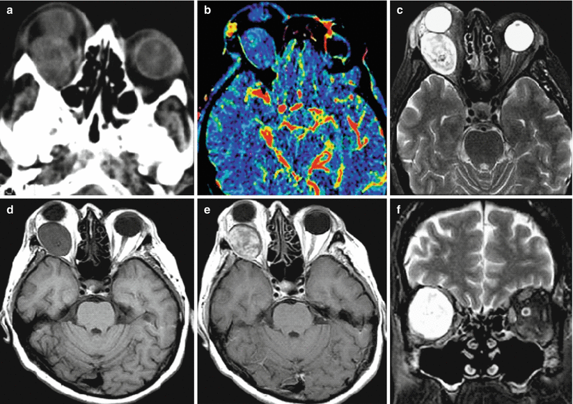

Fig. 37.1
Neurofibroma. On CT (a) in the right orbit, there is a large isodense tumor causing severe exophthalmos. Based on CT perfusion findings (b), hemodynamic parameters in the tumor (CBV) are not increased as compared to the white substance. On T2-weighted (c, f) and T1-weighted MRI (d), an encapsulated tumor is detected with a heterogeneous structure of the MR signal (hyperintense in T2-weighted images and a decreased signal in T1-weighted images). On T1-weighted MRI with contrast enhancement, there is intense, uneven accumulation of the contrast agent in the tumor (e)
Neurofibroma malignizes less often than neuroma. Malignization usually occurs in patients with type I neurofibromatosis. In the case of malignization, they become more aggressive and may grow intracranially.
37.2 Glioma of the Optic Nerve
Glioma of the optic nerve is often a benign, slow-growing tumor, amounting to 66% of all primary tumors of the optic nerve. In 65% of cases, glioma develops within the first decade of life, but only 5% of gliomas are detected before 2 years of age. According to Shields (1998), gliomas account for 1–2% of all orbital tumors, while according to Baert (2006), up to 4%. Women are affected more often than men (3:2). 30–40% of patients with gliomas have manifestations of NF I, and 15% of patients with NF I have an optic nerve glioma.
Stay updated, free articles. Join our Telegram channel

Full access? Get Clinical Tree


