

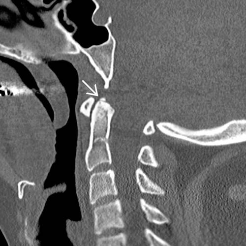
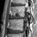
 located adjacent to the odontoid tip. The ossicle is in orthotopic position, and CVJ alignment is normal.
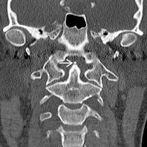
 . Note that there is minimal flattening of the odontoid tip adjacent to the ossicle, supporting classification as a persistent terminal odontoid ossicle.
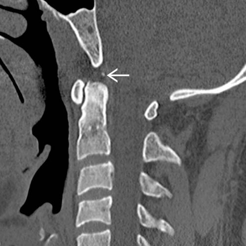
 positioned between the odontoid tip and clivus. The odontoid tip has remodeled.
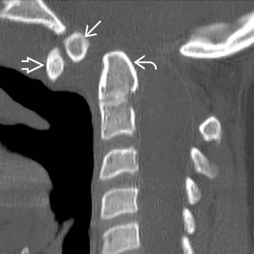
 relative to the nearly normal-sized odontoid process
  . There is a large dystopic ossiculum terminale
  . Although debatable, the nearly normal size of the odontoid process favors classification as an ossiculum terminale rather than os odontoideum.
IMAGING
General Features
• Best diagnostic clue
 Separate ossicle with intact cortical margin positioned above normal-sized odontoid process in adolescent or adult


Separate ossicle with intact cortical margin positioned above normal-sized odontoid process in adolescent or adultRelated posts:

Stay updated, free articles. Join our Telegram channel
Full access? Get Clinical Tree
