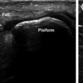
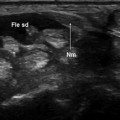
Fig. 16.1
Trapeziometacarpal osteoarthritis. Radiography (a) shows narrowing of the joint space and subchondral bone sclerosis. Ultrasonography confirms the presence of both (b) and also reveals synovial hypertrophy and a small osteophyte (c)
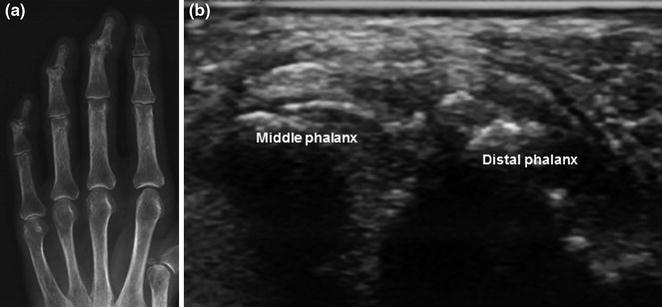
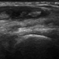
Fig. 16.2
Osteoarthritis of the distal interphalangeal joints. The radiographic examination (a) reveals joint space narrowing, subchondral sclerosis, and subluxation at the levels of the middle, ring, and little fingers. Sonography (b) confirms the reduced joint space and subchondral sclerosis and reveals synovial hypertrophy
Radiography (Figs. 16.1a and 16.2


Related posts:
Stay updated, free articles. Join our Telegram channel
Full access? Get Clinical Tree
