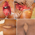
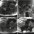
Fig. 9.1
Osteoblastoma of the sacrum in a 19-year-old male. (a–b) CT scan axial images show that the lesion is well limited and is surrounded by reactive sclerosis. (c) CT-3D reconstruction shows the anatomic relationship with vascular structures. (d–e) Intraoperative pictures. A cryosurgery has been performed as local adjuvant. (f) Specimen after curettage. Note the “ice balls”
9.5.3 Sacral Resection
9.5.4 Radio-Frequency Thermal Ablation
Image-guided radio-frequency thermal ablation (RFTA) reduces the pain and improves the function and quality of life of patients with painful bone tumors. It has been used to treat benign bone tumors and tumor-like lesions, and it is the gold-standard procedure for most osteoid osteomas [53]. Even if osteoblastoma is histologically similar to osteoid osteoma, this tumor can exhibit aggressive behavior, and recurrences are not uncommon after classic treatment by surgical excision or curettage [54]. RFTA could be performed in the same way as for osteoid osteomas, using electrodes with a longer active tip or a greater number of ablation sessions or multi-tip array probes [55].
9.6 Oncologic Outcome
In our literature review, we were able to find only one case of sacral osteoblastoma that died of disease [17]. Dal Cin et al. reported a 35-year-old man with osteoblastoma of the sacroiliac joint treated with partial resection. The pathologic examination revealed an intralesional margin, and the patient was subsequently treated with chemotherapy and radiation therapy (without effect) and died 2 years after diagnosis [17]. The authors explained the aggressive clinical behavior with a possible malignant transformation of the stromal component [17].
The recurrence rates for osteoblastomas of the spine after intralesional excision have been reported between 10 and 19% [3, 56, 57], but the rate approaches 50% in the more aggressive pattern. Some studies reported a correlation between the incidence of recurrence and a previous inadequate treatment in non-referral centers [52, 58]. Boriani et al. [58], in a series of 50 osteoblastomas of the mobile spine, reported an incidence of recurrence among “not intact” patients (cases already treated elsewhere) significantly higher compared to “intact” patients (cases treated in the same institution since the beginning): 50% vs. 5%. Some tumors are more prone to recurrence than others, and some factors relating to surgery may explain this, but the molecular features driving this for osteoblastoma are not known. Also in authors’ experience, previous inadequate intralesional surgery was associated with higher rate of local recurrence (40%—two local recurrences in five cases) than patients primarily treated at a specialized tumor center with high levels of surgical and oncologic expertise (7.7%—one local recurrence in 13 cases) [6].
Conflict-of-Interest Statement
No benefits have been or will be received from a commercial party related directly or indirectly to the subject matter of this article.
References
1.
Unni KK. Benign osteoblastoma (giant osteoid osteoma). In: Unni KK, editor. Dahlin’s bone tumours: general aspects and data on 11087 cases. 5th ed. Philadelphia: Lippincott-Raven; 1996. p. 131–42.
2.
Greenspan A, Remagen W. Differential diagnosis of tumors and tumor-like lesions of bone and joints. Philadelphia: Lippincott-Raven; 1998. p. 25–366.
3.
4.
Campanacci L. Osteoblastoma. In: Picci P, et al., editors. Atlas of musculoskeletal tumors and tumor like lesions. Switzerland: Springer; 2014. p. 81–4.CrossRef
5.
Campanacci M. Bone and soft tissue tumors. New York: Springer; 1993.
6.
7.
Bertoni F, Bacchini P, Donati D, Martini A, Picci P, Campanacci M. Osteoblastoma-like osteosarcoma. The Rizzoli Institute experience. Mod Pathol. 1993 Nov;6(6):707–16.PubMed
8.
Dorfman HD, Weiss SW. Borderline osteoblastic tumors: problems in the differential diagnosis of aggressive osteoblastoma and low-grade osteosarcoma. Semin Diagn Pathol. 1984;1:215–34.PubMed
9.
Biagini R, Orsini U, Demitri S, et al. Osteoid osteoma and osteoblastoma of the sacrum. Orthopedics. 2001;24(11):1061–4.PubMed
10.
Bessou P, Lefournier V, Ramoul A, Vasdev A, Boubagra K, Crouzet G. Benign vertebral osteoblastoma. Report of 6 cases. J Neuroradiol. 1998;25(1):21–31.PubMed
11.
Ozaki T, Liljengvist U, Hillmann A, et al. Osteoid osteoma and osteoblastoma of the spine: experiences with 22 patients. Clin Orthop Relat Res. 2002;397:394–402.CrossRef
12.
13.
14.
15.
Khan IS, Thakur JD, Chittiboina P, Nanda A. Large sacral osteoblastoma: a case report and review of multi-disciplinary management strategies. J La State Med Soc. 2012;164(5):251–5.PubMed
Related posts:


Stay updated, free articles. Join our Telegram channel

Full access? Get Clinical Tree


