© Springer International Publishing AG 2017
Pietro Ruggieri, Andrea Angelini, Daniel Vanel and Piero Picci (eds.)Tumors of the Sacrum10.1007/978-3-319-51202-0_1616. Osteosarcoma of the Sacrum
(1)
Department of Orthopaedics and Orthopedic Oncology, University of Padova, Padova, Italy
(2)
Department of Surgery, University of South Florida, H. Lee Moffitt Cancer Center, Tampa, FL, USA
16.1 Introduction
Osteosarcoma and Ewing’s sarcoma are the most common primary malignant bone tumors in childhood and adolescence, most occurring during the first two decades [1, 2]. Osteosarcoma rarely involves the spine (1–3% of all osteosarcomas), and the sacrum is one of the most common spinal locations [2–9]. Osteosarcoma is the third most frequent primary malignant tumor of the sacrum after chordoma and Ewing’s sarcoma. Secondary osteosarcoma may occur in patients that received pelvic radiation treatment or as sarcomatous degeneration in patients with polyostotic Paget’s disease [10–15].
There have been several articles on the treatment of patients with osteosarcoma of the spine [4–7, 16–32]; however, those publications included case reports or selected patients from small series, making the optimal evidence-based therapeutic approach very difficult. Wuisman et al. [23] reported a case of sacral osteosarcoma and performed a review of all reported cases since 1984–2001: they were able to find only 11 patients [7, 18, 25–32]. In the last 15 years, still some data appear in the literature, indicating that the combination of chemotherapy with adequate surgical procedures (hemi/total sacrectomies) may increase survival in this poor prognostic tumor site [4, 20–22, 24].
16.2 Epidemiology, Presentation, and Diagnosis
Patients with primary spinal osteosarcoma are older than those with osteosarcoma of the extremity (mean age of about 38 years old) [5, 6, 8, 33]. Exposure to radiation is a proven exogenous risk factor for secondary osteosarcoma of the bone. Incidence of sarcomas postirradiation therapy comprises about 0.1% of all cancer cases, and the sarcomas usually appear 10–20 years posttreatment (thus radiation-induced sarcoma is typical of adult age) [10–15]. It has been demonstrated that osteosarcoma could be related to pelvic radiation therapy [10, 13–14].
As well as for other sacral tumors, early diagnosis is difficult and pain and swelling are the most frequent symptoms of sacral osteosarcoma. Neurological symptoms are associated with large tumors. A detailed history with a complete physical exam should be performed prior to any evaluation. Ozaki et al. reported in 2002 their experience in 15 osteosarcomas of the sacrum and 7 of the mobile spine [4]: the duration of symptoms between onset and diagnosis ranged from 2 to 18 months, and the most common was represented by pain (almost all) followed by neurological disorder (half of the cases). The median tumor size in sacral tumors was 8.0 cm. Other reports were results in terms of diagnostic delay and tumor size [8, 34]. The laboratory findings may show an increase in alkaline phosphatase (AP) and lactic dehydrogenase in the serum (about 30% of cases) [35–38]. In some cases mild anemia and high erythrocyte sedimentation rate may also be present at diagnosis [35, 36].
16.3 Imaging
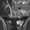
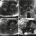
Most spinal osteosarcomas are pathologically osteoblastic, resulting in typical mineralized lesions on radiographs and CT, even if rarely osteolytic lesions also occurred [4, 29, 37, 39]. Computed tomography (CT), magnetic resonance imaging (MRI), angiography, and dynamic bone scintigraphy are used to evaluate the extension of tumors and the involvement of surrounding structures such as vessels, nerves, and soft tissues [40, 41]. CT of the lung is part of the basal staging. MRI is also useful in the assessment of patients with intraosseous tumor spread, particularly in the sacrum or sacroiliac joint, or in the identification of neural compression [8].
Nuclear medicine imaging techniques (bone scans and PET/CT) are being used increasingly to aid in the initial staging/metastatic evaluation and response to therapy. Isotope scans with technetium [41] or thallium [42] are a standard part of the staging because they show the intense hotspot of the tumor and are very sensitive in detecting any skip or distant bony metastases [43]. 18-Fluorodeoxy-glucose positron emission tomography (18FDG-PET) combined with a whole-body CT is being increasingly used in staging and also in treatment monitoring [44–46]. On 18FDG-PET/CT, there is increased uptake within the tumor, which decreases following effective neoadjuvant therapy [44–46]. Whole-body MRI and PET/CT are currently being evaluated for the detection of metastatic disease [47].
Biopsy is a key diagnostic method for sacral osteosarcomas and should be carefully planned according to the definitive surgery, in order to avoid improper treatments or negative effects on survival [48–50]. Conventional high-grade osteosarcoma is the most frequent variant at histopathologic evaluation. Multicentric osteosarcoma is a rare type of the disease characterized by a synchronous or metachronous appearance of multiple skeletal lesions. Some cases with sacral involvement have been reported [51, 52] with extremely poor prognosis. On the other hand, low-grade osteosarcoma of the sacrum (well-differentiated intraosseous osteosarcoma) has been described in only one report in literature [53].
16.4 Treatment
In recent years, multidisciplinary approach and aggressive adjuvant/neoadjuvant chemotherapy have increased the oncologic outcome of patients with osteosarcoma [5, 6, 35, 37, 54–60], even if spinal involvement has been linked with a very poor prognostic outlook with median survival times of only 10–23 months. Much like with Ewing’s sarcoma, a combination of chemotherapy with surgery (when possible) is also the standard therapy in tumors involving the axial skeleton. Some authors suggested that patients with osteosarcoma of the spine should be treated with a combination of chemotherapy and at least marginal surgery [4]. Postoperative radiotherapy can also be applied in the treatment program and may be of benefit in selected patients.
16.4.1 Chemotherapy
Currently, chemotherapy is undoubtedly the method that is likely to cure the greatest proportion of patients with osteosarcoma. However the association with surgery is essential for the local control and the management program of all patients. In fact, in the presence of effective chemotherapy, osteosarcoma is rarely cured without surgical resection [5, 17, 61]. Doxorubicin, cisplatin, high-dose methotrexate, ifosfamide, and etoposide have antitumor activity in osteosarcoma and are frequently used with different protocols as the basis of treatment [54, 57–59, 62]. The selection of postoperative adjuvant chemotherapy based on the degree of the tumor necrosis induced by preoperative therapy improves the patient survival rate [59, 62]. New drugs such as bisphosphonates, interferon, interleukin, and monoclonal antibodies have been trialed in preclinical and clinical studies, showing encouraging results [59]. Specific aspects on protocol of treatment are analyzed in the dedicated chapter.
16.4.2 Sacrectomy
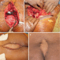
Recent progression of surgical techniques may enable total sacrectomy to improve the survival of patients [63–68]. Patients whose tumors can be completely resected with adequate margins should be approached with curative intent. Simon et al. [29] reported a patient disease-free almost 5 years after surgical excision alone. However, the surgical intervention is quite challenging given the magnitude of treatment, the significant compromise of neurological status, and the high risk of complications [69–72]. To date, the question whether this type of surgery is really beneficial to the affected patients is still debated. Ozaki et al. [4] show that complete tumor resection may improve their prognosis. In the author’s experience, most patients have unresectable or partially resectable tumors, or metastases at presentation, and thus are not good candidates for a surgical treatment (or adequate margins cannot be achieved). Obviously sacral resection in low-grade tumors should be considered the mainstay of treatment and has been successfully reported [53].
There is no absolute contraindication for surgical resection because the decision is dictated by local practice and the surgical expertise of the tumor center. Relative contraindications for resection are large extraosseous extension, major neurovascular involvement, high mortality/morbidity risk with extensive surgery, and unavailability of experienced multidisciplinary team.
16.5 Radiation Therapy
In general, radiotherapy has a limited role in the management of osteosarcoma because of the relative radioresistance and the need for a large dose of radiation to achieve clinical response, but there are anatomical locations in which the possibility of complete surgical resection is unfeasible [73–75]. However, the effect of chemotherapy alone is usually temporary, and there is a need of intensive treatment for local control. In these cases, radiation therapy may be an option to try to extend the progression-free interval. Another possible scenario is the use of adjunctive irradiation in patients who underwent intralesional or inadequate surgery, in which the overall survival was better compared with patients that not received further local treatments [4, 6].
Radiotherapy may provide significant palliation in patients with unresectable sacral osteosarcomas (or patients that refused surgery), even if some cases successfully treated with combination of chemotherapy and radiation have been reported in literature [6, 59, 76, 77].
Considering that high-dose conventional radiation cannot usually be given in the sacrum and new radiation therapy techniques (e.g., proton beam and heavy ion carbon therapy) are available, the role of radiation therapy in osteosarcoma may need to be reinvestigated with modern techniques that may extend indications [78]. Targeted internal radiotherapy with Sm-153-EDTMP could be an additional treatment option for some patients with inoperable tumors [79, 80].
16.6 Oncologic Outcome
Although multimodal treatment, osteosarcoma of the sacrum has a significantly worse outcome than it affects other sites [5, 6, 35, 58, 59]. In the recent ESMO guidelines, primary metastases, axial or proximal extremity tumor site, large tumor size, elevated serum AP or LDH, and older age are considered adverse prognostic or predictive factors [58].
The median survival for patients with osteosarcoma of the spine has been reported in the range of 6–10 months [5–7, 16]. Shives et al. [6] reported that only 7 of 26 patients treated for their spinal osteosarcomas up to 1980 survived for 1 year. In a large retrospective series of spinal osteosarcomas (15 with tumors of the sacrum and 7 with tumors at other sites), 86% of the patients survived 1 year, but only 3 were alive at 6 years of follow-up [4]. Li et al. [20] reported a series including two cases of osteosarcoma of the sacrum treated with hemisacrectomy and adjuvant chemotherapy: one was alive with disease with local recurrence after resection with marginal margins at 49 months of follow-up, whereas the other was alive with no evidence of disease at 24 months (wide margin). Guo et al. [21] reported a series on en bloc sacrectomies including two patients affected by osteosarcoma: one was alive with disease with local recurrence at 11 months of follow-up and the other alive with no evidence of disease at 20 months. Arkader et al. [22] reported two cases of sacral osteosarcoma in pediatric age, disease-free at 8 years of follow-up after sacrectomy and chemotherapy. Sundaresan et al. [7] suggested that the combination of surgery, chemotherapy, and radiation may increase survival, showing three patients who survived longer than 36 months without evidence of disease.
Conflict-of-Interest Statement
No benefits have been or will be received from a commercial party related directly or indirectly to the subject matter of this article.
References
1.
2.
Unni KK. Dahlin’s bone tumors general aspects and data on 11,087 cases. 5th ed. Philadelphia: Lippincott-Raven; 1996.
4.
5.
Barwick KW, Huvos AG, Smith J. Primary osteogenic sarcoma of the vertebral column: a clinicopathologic correlation of ten patients. Cancer. 1980;46:595–604.PubMed
Stay updated, free articles. Join our Telegram channel

Full access? Get Clinical Tree


