Osteosarcoma—Vertebral


Clinical Presentation


The patient is a 23-year-old man who presented with severe progressive neck pain, left arm pain, and symptoms of cord compression. The patient had received a course of radiotherapy 8 years previously for a malignant neck soft tissue tumor.


Imaging Presentation


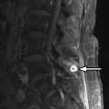
Computed tomography (CT) images reveal bone destruction of the C3 and C4 vertebral bodies and near complete collapse of the C4 vertebral body. There is a large area of tumoral bone formation along the left anterolateral aspect of the C4 vertebral body ( Fig. 57-1 ) . Magnetic resonance (MR) imaging revealed heterogeneous enhancement of a large paraspinal mass at C3-4 on the left with encasement of the left vertebral artery ( Figs. 57-2 to 57-4 ) . There was enhancing tumor extending into the left C3-4 neural foramen and into the epidural space anteriorly and on the left (see Fig. 57-4 ). The spinal cord was compressed from the left and displaced posteriorly and to the right by the epidural tumor (see Figs. 57-3 and 52-4 ). Surgery was performed to decompress the spinal cord and remove the tumor, which proved to be osteosarcoma. Because of the history of radiation therapy, this was presumed to be a radiation-induced vertebral osteosarcoma.


Figure 57-1


Radiation Induced Cervical Vertebral Osteosarcoma.

Axial CT image, C4 level. Heterogeneous bone destruction resulting in vertebral collapse and tumoral bone formation ( short thick arrows ) projects anterolateral to the vertebral body on the left. Heterogeneous epidural tumor ( long thin arrows ) displaces the cord posteriorly to the right.


Figure 57-2


Radiation Induced Cervical Vertebral Osteosarcoma.

Same patient as in Figure 57-1 . Sagittal fat-saturated T2-weighted image. Tumor has destroyed the C4 and C5 vertebral bodies. The C4 vertebral body has collapsed resulting in C4 vertebral planum ( black arrowhead ). C4-5 ventral epidural tumor displaces the cord posteriorly. Prevertebral soft tissue mass ( arrows ) extends from C2 to C5 level. C5 = C5 vertebral body.


Figure 57-3


Radiation Induced Cervical Vertebral Osteosarcoma.

Same patient as in Figures 57-1 and 57-2 . Sagittal unenhanced T1-weighted image A and contrast-enhanced T1-weighted image B . The prevertebral mass ( white arrows ), adjacent infiltrated vertebrae and anterior epidural mass ( black arrows ) enhance following IV contrast. Collapsed, posteriorly displaced C4 vertebral body ( black arrow in A , arrowhead in B ) and epidural tumor ( black arrows in image B ) displace the spinal cord posteriorly. C5 = C5 vertebra body.


Figure 57-4


Radiation Induced Cervical Vertebral Osteosarcoma.

Same patient as in Figures 57-1 to 57-3 . Axial unenhanced MR image A and contrast-enhanced image B , at C3-4 level. The patient’s neck is turned toward the right. Heterogeneously enhancing mass encases the left vertebral artery ( VA ) and extends into the C3-4 foraminal and paraspinal soft tissues on the left ( arrows ). The epidural tumor on the left displaces the spinal cord ( C ) to the right of midline.


Discussion


Osteosarcoma, also known as osteogenic sarcoma , is a highly malignant primary tumor of bone that produces immature osteoid matrix. No more than 3% of osteosarcomas originate in the spine, so these are very uncommon vertebral tumors. Osteosarcomas represent up to 5% of primary malignant spine tumors, vertebral myeloma/plasmacytoma, and lymphoma being far more common. Most vertebral osteosarcomas manifest in the second through fifth decades of life. The much more common appendicular skeleton osteosarcomas usually occur in children or adolescents. Most primary vertebral osteosarcomas arise in the vertebral body but often infiltrate the pedicles and can involve the posterior arch. Only 10% to 17% of osteosarcomas are reported to occur primarily in the posterior vertebral arch. Primary vertebral osteosarcomas most commonly occur in the lumbar spine, followed by the thoracic spine, sacrum, and least commonly in the cervical spine. Most primary vertebral osteosarcomas have a bulky, lobulated soft tissue component that breaks through the cortex and commonly extends into the spinal canal or paraspinal soft tissues (see Figs. 57-1 to 57-4 ).


The majority of vertebral osteosarcomas are metastatic tumors that originate from primary osteosarcomas in the appendicular skeleton or adjacent soft tissues of the extremities in children, adolescents, or young adults ( Figs. 57-5 to 57-7 ) . These patients often have hepatic and pulmonary metastases in addition to vertebral metastases.


Figure 57-5


Metastatic Osteosarcoma to T12 Vertebral Body from Lower Extremity Primary Osteosarcoma.

Axial CT image shows sclerotic metastasis ( arrow ) in T12 vertebral body. This may be mistaken for a bone island or osteoblastic metastasis from another type of primary tumor.


Figure 57-6


Metastatic Osteosarcoma to T12 Vertebral Body.

Sagittal MR images in same patient as in Figure 57-5 . A well-defined sclerotic osteosarcoma metastasis ( arrow ) is T2 hypointense in image A and T1 hypointense in image B and enhances slightly so that it is nearly isointense relative to adjacent marrow on contrast enhanced image C . Bone islands may have a similar CT, T1, and T2 MR appearance, but bone islands do not enhance following IV contrast.

Only gold members can continue reading. Log In or Register to continue

Stay updated, free articles. Join our Telegram channel

Aug 25, 2019 | Posted by in NEUROLOGICAL IMAGING | Comments Off on Osteosarcoma—Vertebral

Full access? Get Clinical Tree

Get Clinical Tree app for offline access