Ovarian Torsion
Claire S. Cooney
Ovarian torsion is the twisting of the ovary on its ligamentous attachments causing vascular compromise. It is an uncommon event, but accounts for 3% of all gynecologic operative emergencies and can have significant adverse sequelae. Symptoms can be vague, and therefore imaging is crucial for prompt diagnosis and treatment to avoid irreparable damage to the ovary.
Etiology.
Twisting of the ovarian pedicle leads to compromise of lymphatic and venous drainage, causing the ovary to become enlarged and edematous. Next, ovarian ischemia can occur due to compromise of arterial flow, resulting in infarction, necrosis, and hemorrhage. Risk factors include an ovarian mass, pregnancy, and ovarian hyperstimulation syndrome.
Symptoms and Signs.
The classic clinical scenario is a woman of childbearing age who presents with acute onset of lower abdominal pain associated with nausea and vomiting. The clinical presentation can include one or more of the following:
Unilateral pelvic or lower abdominal pain (right more common than left), possibly intermittent, and usually acute in onset (in contrast to slowly developing migratory pain of appendicitis).
Nausea and vomiting can occur during episodes of pain.
Peritoneal signs.
Ascites—hemorrhagic, inflammatory, or transudative.
Fever—rare and may be due to ovarian necrosis.
Clinical workup must include a careful pelvic examination before imaging, as the presence of an adnexal mass dramatically increases possibility of torsion. Β- human chorionic gonadotropin (Β-hCG) is required for premenopausal patients.
Differential Diagnosis.
If the Β-hCG is positive, ectopic pregnancy must be considered. If it is negative, appendicitis, pelvic inflammatory disease, tubo-ovarian abscess, endometriosis, degenerating fibroid, hemorrhagic cyst, gastroenteritis, intussusception, diverticulitis, and menstrual cramping are in the differential diagnosis.
Indications.
Ultrasonography is the examination of choice for the diagnosis of ovarian torsion because it can accurately and rapidly differentiate ovarian torsion from other pelvic disorders that have the same nonspecific clinical presentation.
Protocol.
Transabdominal scan with full bladder, then endovaginal examination with color Doppler after bladder is emptied. A Foley catheter may be placed to facilitate adequate bladder filling. Both adnexa should be examined.
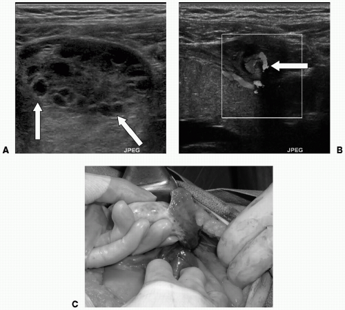
 Figure 44-1. A:
Get Clinical Tree app for offline access
Stay updated, free articles. Join our Telegram channel
Full access? Get Clinical Tree


|