Figure 28.1.1
Simple ovarian cyst. A: Coronal (COR RO) and (B) sagittal (SAG RO) images of the right ovary demonstrating a simple ovarian cyst (arrows). Normal ovarian tissue (arrowheads) is seen around a portion of the cyst.
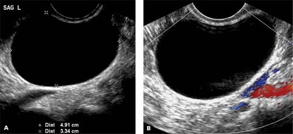
Figure 28.1.2
Simple ovarian cyst. A: Sagittal image of left ovary (SAG L) demonstrating a simple ovarian cyst (calipers) measuring 4.9 x 3.3 cm. B: Color Doppler image of cyst showing no internal blood flow nor flow in the thin, smooth wall of the cyst.
28.2. Hemorrhagic Ovarian Cysts
Description and Clinical Features
Occasionally, a functional ovarian cyst bleeds into itself. When this happens, the cyst is termed hemorrhagic. Hemorrhagic cysts, like simple ovarian cysts, are benign lesions of the ovary that most often resolve spontaneously, without requiring surgical intervention. Occasionally, acute hemorrhage into a cyst will cause sudden onset of pelvic pain, and, rarely, a hemorrhagic cyst ruptures.
Hemorrhagic cysts typically occur in women of reproductive age and not in postmenopausal women.
Sonography
Hemorrhagic cysts appear as complex ovarian lesions, often with fine septations that form a reticular pattern throughout the cyst (Figures 28.2.1 and 28.2.2). The pattern of septations is sometimes described as weblike or lacy. The fluid within the cyst often has scattered echoes. The walls of a hemorrhagic cyst may be thin and smooth, or they may be focally or diffusely thickened. No blood flow should be seen within the cyst or its septations, but flow may be seen in its walls (Figure 28.2.2).
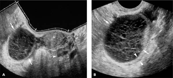
Figure 28.2.1
Hemorrhagic ovarian cyst. A: Transvaginal panoramic view demonstrating complex right ovarian cyst (arrows) adjacent to a normal uterus (U) and left ovary (arrowhead). B: Magnified view of right hemorrhagic ovarian cyst (arrows) showing internal septations forming a reticular pattern throughout the cyst. The cyst wall is slightly thickened on one side (arrowheads).
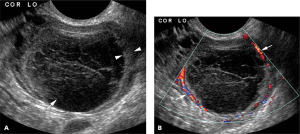
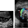
Figure 28.2.2
Hemorrhagic cyst with blood flow in the wall. A: Coronal image of left ovary (COR LO) demonstrating a hemorrhagic cyst with echoes in the fluid of the cyst (arrow) and focal thickening of the wall (arrowheads). B: Color Doppler image showing circumferential flow in the cyst wall (arrows) and no flow in the septations or echoes within the cyst.
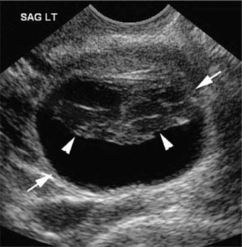
Figure 28.2.3
Hemorrhagic cyst with retracted clot. Sagittal image of left ovary (SAG LT) showing a cyst (arrows) with septations and solid-appearing component anterior (arrowheads) and anechoic fluid posterior.
The appearance of a hemorrhage cyst typically evolves after the initial hemorrhage. With time, after the initial reticular pattern, the echoes within the cyst may conglomerate to one side of the cyst (Figure 28.2.3), sometimes with a concave contour. The conglomeration represents retracting clot. No blood flood should be seen within this conglomeration. With continued involution, the cyst decreases in size and the septations and conglomeration of echoes become smaller or disappear (Figure 28.2.4).
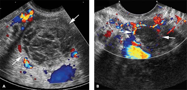
Figure 28.2.4
Evolving hemorrhagic cyst. A: Image of ovary demonstrating a hemorrhagic cyst (arrows) with septations, echoes in the fluid, and blood flow in the wall. B: On follow-up scan 6 weeks later, the hemorrhagic cyst (arrows) is smaller, there are fewer septations, and the septations are thinner.
28.3. Ovarian Teratomas
Description and Clinical Features
The most common benign neoplasm of the ovary is a dermoid cyst, also called a mature cystic teratoma. This tumor is of germ cell origin and is often found in women of reproductive age. It is bilateral in 10%–15% of cases. Most dermoid tumors are asymptomatic, but occasionally they cause lower abdominal pain, swelling, and irregular menses. Ovaries containing dermoids are at risk of torsion. Surgical excision is the usual treatment of these lesions. At pathology, the tumor may be found to contain fat and sometimes bone, teeth, or hair. Rarely, an ovarian teratoma is malignant.
Sonography
Mature cystic teratomas of the ovary often have a typical sonographic appearance that allows them to be differentiated from other ovarian neoplasms. The characteristic appearance is a complex, partially cystic mass in the ovary that contains one or more highly echogenic regions that may shadow (Figure 28.3.1). Echogenic lines and dots (Figure 28.3.2) may be seen within the cystic areas of the mass, representing hair. Some of the highly echogenic regions represent fat within the tumor. The fat may be seen floating on top of other fluid in the lesion, leading to the sonographic finding of a fluid–fluid level (Figure 28.3.3). Other echogenic regions with shadowing may contain solid nodules of tissue in the wall of the dermoid (Figure 28.3.4) or densely calcified structures representing teeth or bone. Dermoid tumors typically have little or no internal flow on color Doppler imaging.
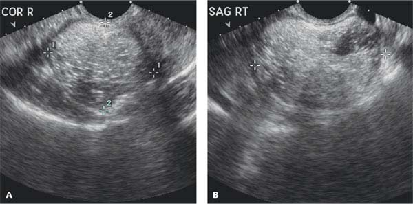
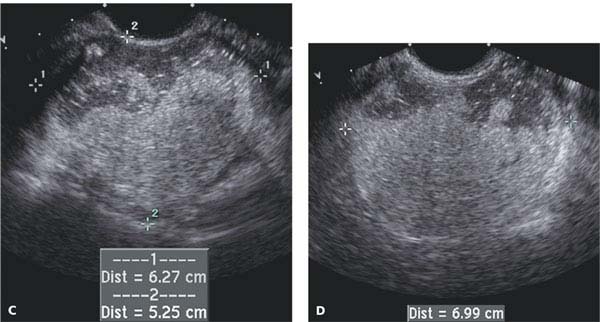
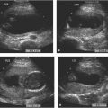
Figure 28.3.1
Ovarian dermoid tumors containing fat. A: Coronal (COR R) and (B) sagittal (SAG RT) transvaginal images of mass (calipers) in right ovary filled with complex highly echogenic material representing fat. C: Coronal and (D) sagittal images of large mass (calipers) with lobular regions of increased echogenicity representing fat and regions of decreased echogenicity representing fluid.
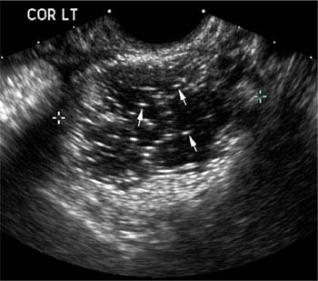
Figure 28.3.2
Ovarian dermoid tumor with echogenic lines and dots. Coronal image of left ovary (COR LT) demonstrating a dermoid tumor (calipers) with echogenic short lines and dots (arrows) within the cystic portion.
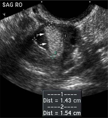
Figure 28.3.3
Ovarian dermoid tumor with fluid–fluid level. Sagittal image of right ovary (SAG RO) with complex lesion (calipers) containing hypoechoic and echogenic regions that form a linear interface (arrows) due to layering of fat and other fluid.
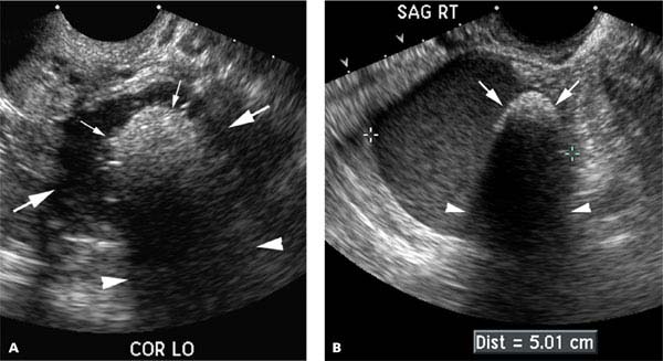
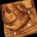
Figure 28.3.4
Ovarian dermoid tumor with highly echogenic solid nodule that shadows. A: Transvaginal image of ovary demonstrating complex mass (large arrows) with an echogenic solid nodule of tissue (small arrows) protruding into the mass and causing acoustic shadowing (arrowheads) behind the solid nodule. B: Sagittal image in another patient showing large dermoid (calipers) that is predominantly cystic but has an echogenic solid nodule (arrows) protruding from the wall and causing acoustic shadowing (arrowheads).
28.4. Ovarian Benign Neoplasms Other Than Teratomas
Description and Clinical Features
Ovarian neoplasms that arise from epithelial cells and surrounding stromal cells can be malignant or benign. The most common benign neoplasms are mucinous or serous cystadenomas. Less common benign tumors arising from these cells include transitional cell (Brenner) tumors. Benign ovarian neoplasms can also arise from granulosa, theca, and Sertoli and Leydig cells. These neoplasms include ovarian fibromas, granulosa cell tumors, thecomas, and Sertoli–Leydig cell tumors.
The distinction between benign and malignant ovarian neoplasms cannot be made with certainty based on clinical presentation or imaging findings.
Sonography
Serous and mucinous cystadenomas of the ovary are typically complex ovarian lesions with septations separating areas of anechoic fluid (Figure 28.4.1). Blood flow can often be identified in the septations with color Doppler. Mucinous cystadenomas tend to have more septations than serous cystadenomas and sometimes the fluid in mucinous cystadenomas contains low-level echoes (Figure 28.4.2). Some benign tumors contain both anechoic cystic regions and solid or complex cystic areas (Figure 28.4.3). Occasionally, solid tumor nodules, containing vessels visible with color Doppler, are seen in the wall of the neoplasm, although this finding is more frequently seen with malignant than benign neoplasms.
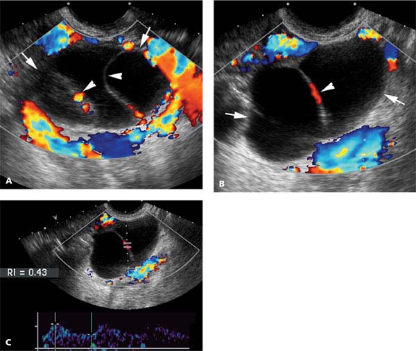
Figure 28.4.1
Ovarian serous cystadenoma. A and B: Transvaginal color Doppler images of cystic ovarian lesion (arrows) with anechoic fluid and a few thin septations (arrowheads). Color Doppler shows blood flow within the septations. C: Doppler gate placed on blood flow in a septation demonstrates an arterial waveform (calipers) with a resistive index (RI) of 0.43.
Related posts:


Stay updated, free articles. Join our Telegram channel

Full access? Get Clinical Tree


