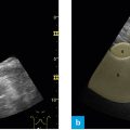
Fig. 1: Position of the probe, subxiphoid and transverse – pancreas.
From its longitudinal position the probe is rotated 90° in counterclockwise direction with the superior mesenteric artery (SMA) in the center of the image. One continues to see the SMA in the center of the image on the cross-section. The pancreas can be identified by its surrounding structures. The probe marker points to the investigator.
The pancreas is usually difficult to visualize on ultrasound. Even in young and healthy persons it is poorly contrasted and therefore not easily located. The pancreas is most reliably identified by its surrounding structures. The best method is to recall the anatomical location of the retroperitoneum on the basis of the aorta or the vena cava, and identify the other visible structures on the basis of these vessels.
Landmark: superior mesenteric artery (SMA)
The following structures should be viewed:
- Superior mesenteric artery (landmark)
- Aorta
- Vena cava
- Splenic vein
- Liver
- Pancreas
- Optional: left splenic vein
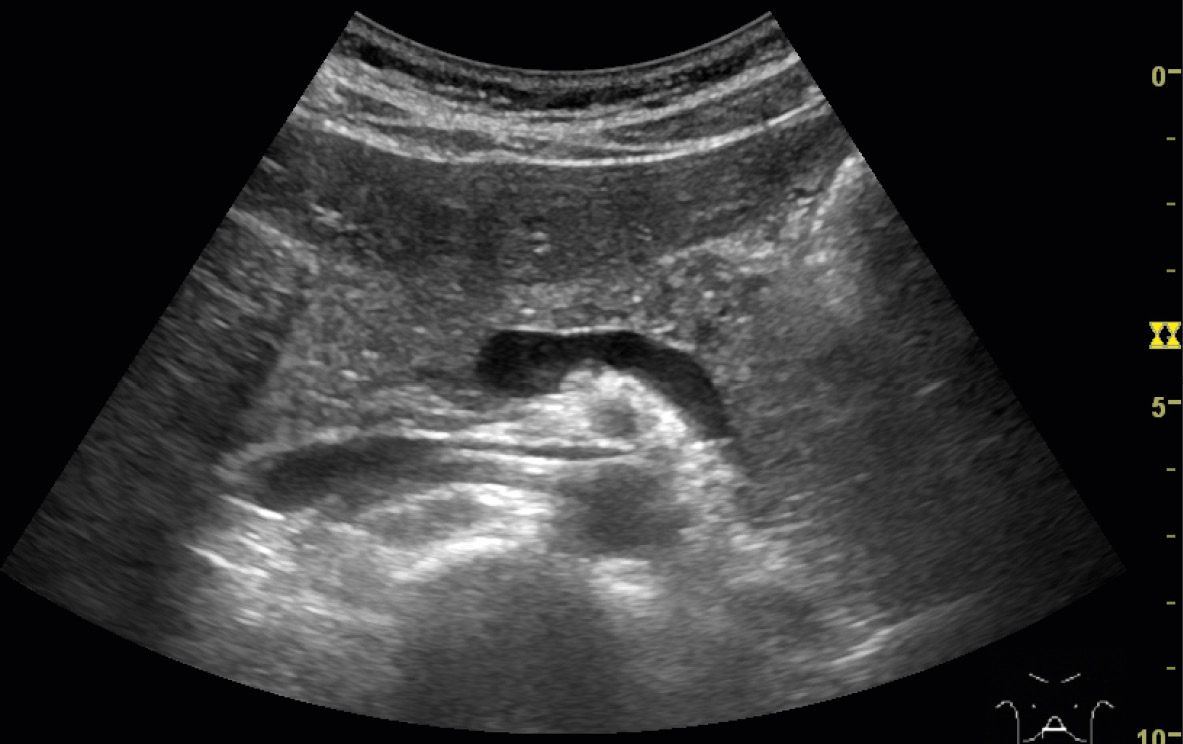
Fig. 2: Ultrasound investigation of the pancreas, B-mode image.
One first identifies the aorta and the vena cava. Another circular hypoechoic structure with a strong hyperechoic frame – the superior mesenteric artery – is seen in the ventral aspect of the aorta. The splenic vein is in the ventral aspect of the superior mesenteric artery. On ultrasound these structures resemble an eye: the superior mesenteric artery is the eye while the splenic vein forms the eyebrow. The liver can also be demarcated clearly by its homogeneous structure. Pancreatic tissue is observed between the liver and the splenic vein. Anatomically the splenic vein is always in the dorsal aspect of the pancreas and therefore seen quite clearly as a dorsal frame of the pancreas. The close proximity of the superior mesenteric artery and the superior mesenteric vein towards the framing head of the pancreas is evident on this section.
Superior mesenteric artery: “the eye”, splenic vein: “the eyebrows”
Related posts:


Stay updated, free articles. Join our Telegram channel

Full access? Get Clinical Tree


