Parallel Imaging
Introduction
Parallel imaging is an approach for reducing scan time and includes techniques such as sensitivity encoding (SENSE) and generalized autocalibrating partially parallel acquisition (GRAPPA). Vendor implementations of these techniques are also known as array spatial and sensitivity encoding technique (ASSET), autocalibrating reconstruction for Cartesian sampling (ARC), integrated parallel acquisition technique (iPAT), and rapid acquisition through parallel imaging design (RAPID).
Vendor Name | Technique | Sensitivity Calibration |
SENSE (Philips) | Prescan | |
Prescan | ||
ARC (GE ) | GRAPPA | Auto |
GRAPPA (Siemens) | GRAPPA | Auto |
mSENSE (Siemens) | Auto | |
RAPID (Hitachi) | Auto |
Parallel imaging requires the use of phasedarray coils. Depending on how many coils are used, time savings (or acceleration factors) of between 2 and 3 are easily achievable, albeit with a similar signal-to-noise ratio (SNR) cost as accompanies other “fast imaging techniques” based on decreasing phase steps or fewer excitations. As a result, parallel imaging is most useful for high SNR situations, for example, CE-MRA and 3T.
Concepts
The “parallel” in parallel imaging refers to the fact that each coil in the phased array receives data at the same time (i.e., in parallel). Recall from Chapter 2 that a phased array is made up of many small coils, with 32 or more available on modern systems.
Parallel imaging works by knowing the local sensitivity of each coil in the phased array. The field of view (FOV) is made intentionally too small in the phase-encoding direction, and the resulting aliasing is unwrapped using this information. The basic concepts were described in the early 1990s, and the first successful clinical implementation was simultaneous acquisition of spatial harmonics (SMASH). SMASH has now been superseded by more sophisticated techniques and the main approaches in clinical use today are SENSE and GRAPPA.
A parallel imaging acquisition with an acceleration factor of 2 acquires data only for every other line of k-space. The corresponding images have a ½ FOV and exhibit aliasing artifact. The SENSE and GRAPPA approaches work either by unwrapping the aliasing in the images or, equivalently, by filling in the missing lines of k-space (Fig. 24-1).
Both approaches make use of the different spatial sensitivities of the coils in the phased array. Due to their distinct spatial locations, each coil has different view of the object (Fig. 24-2). Knowing their spatial locations and sensitivity profiles allows the aliasing to be undone by postprocessing.
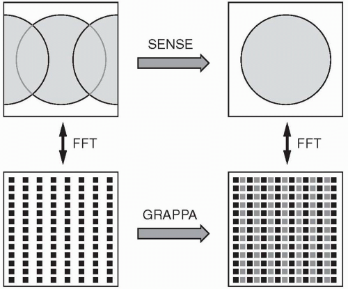 Figure 24-1. Image domain (top) and the associated k-space (bottom), which indicates how aliasing in the image domain is equivalent to undersampling in k-space. The SENSE method unwraps the aliasing in the image, but the same result can be obtained using GRAPPA to fill in lines of k-space. |
 Figure 24-2. Aliased images obtained from an 8-coil phased array. Note each image has the characteristic spatial sensitivity of the coil, such that signals near the coil are strong and those far away are weak. This basic information allows us to work out which signals are in the right location and which signals have been aliased from ½ FOV away. |
Prescan or Autocalibration
There are two common ways to determine the spatial sensitivities of the coils in the phased array. SENSE and GRAPPA differ in the way that spatial sensitivity maps are determined and how the postprocessing is done.
Prescan. A large 3D volume acquisition lasting a few seconds run immediately after the localizers. This gives low-resolution images of the entire region inside the scanner and provides spatial sensitivity maps for all subsequent scans.
Autocalibration. A few extra phase-encoding lines are acquired within each parallel imaging scan. These are used in postprocessing to train the algorithm how to generate missing k-space lines for that specific scan only.
Related posts:
Stay updated, free articles. Join our Telegram channel

Full access? Get Clinical Tree


