and Vincent L. Sorrell2
(1)
Division of Nuclear Medicine and Molecular Imaging Department of Radiology, University of Kentucky, Lexington, Kentucky, USA
(2)
Division of Cardiovascular Medicine Department of Internal Medicine Gill Heart Institute, University of Kentucky, Lexington, Kentucky, USA
Electronic supplementary material
The online version of this chapter (doi: 10.1007/978-3-319-25436-4_5) contains supplementary material, which is available to authorized users.
Normal parathyroid glands are too small to be visualized by routine radionuclide imaging using MPI radiopharmaceuticals. However, benign or malignant enlarged parathyroid glands typically accumulate and retain sufficient activity for imaging detection. Historically, 201Tl chloride was used diagnostically for preoperative identification and localization of culprit glands causing primary and secondary hyperparathyroidism; 99mTc sestamibi is the most widely used radiopharmaceutical today (Coakley et al. 1989; O’Doherty et al. 1992; Smith and Oates 2004b). The hallmark pattern is early uptake with relatively delayed (“differential”) washout from the enlarged parathyroid gland(s) compared to the thyroid gland. However, some lesions may washout more rapidly; since MPI typically commences anywhere from 15 to 90 minutes after injection, be aware that these lesions may appear “warm” rather than the more characteristic “hot” uptake pattern due to the washout kinetics.
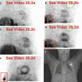
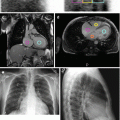
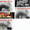
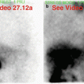
Table 5.1 provides a short list of potential “hot” and “cold” imaging findings related to the parathyroid glands. Particularly if ectopic in location, the enlarged parathyroid gland(s) harboring adenoma, hyperplasia, or carcinoma might be included in the field-of-view during routine MPI (Chamarthy and Travin 2010; Gedik et al. 2007). Figure 5.1 illustrates a “hot” ectopic parathyroid adenoma in the anterior mediastinum just superior to the heart. Mediastinal parathyroid adenomas can be occult during cardiac surgery and may require imaging localization for subsequent directed surgery (Lazar et al. 2005). If large enough, parathyroid lesions can appear “cold” if there is central cystic change or necrosis (Fig. 5.2).
Table 5.1
Differential diagnosis of “hot” and “cold” imaging findings related to the parathyroid glands
Organ system | “Hot” finding | “Cold” finding | References |
|---|---|---|---|
Parathyroid glands | Adenoma Hyperplasia Carcinoma | Cystic adenoma | Chamarthy and Travin (2010) Coakley et al. (1989) Gedik et al. (2007)
Stay updated, free articles. Join our Telegram channel
Full access? Get Clinical Tree
 Get Clinical Tree app for offline access

|

