Chapter 29
Parotid Sialolithiasis
Epidemiology
Parotid sialoliths usually occur between 30 and 50 years of age and are slightly more common in men. Salivary gland calculi in children are rare, and when present, usually involves the submandibular gland. Between 10 and 20% of salivary gland stones occur within the parotid gland. Two thirds of parotid stones are single, whereas one third are multiple. Bilateral stones are unusual unless associated with an autoimmune sialadenitis. Multiple punctate calculi are indicative of sialadenitis.
Clinical Findings
Patients often present with recurrent episodes of pain and swelling of the parotid region, usually associated with eating. The symptoms usually last between 2 and 3 hours and gradually subside. Larger stones may completely obstruct the gland resulting in secondary infection and chronic atrophy if left untreated.
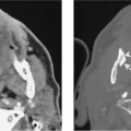
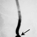
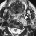
Symptomatic stones are usually found in Stensen’s duct. A patient may have multiple intraparotid stones but only become symptomatic when a stone migrates and obstructs Stensen’s duct. Strictures may result from calculi with or without an underlying infection.
Pathology
Parotid sialoliths consist of calcium phosphate in the form of hydroxyapatite with small amounts of magnesium, carbonate, and ammonium. The matrix consists of carbohydrates and amino acids.
Treatment