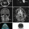
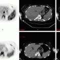
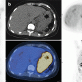
Fig. 2.1
Lymphocyte predominant (LP) cells in nodular lymphocyte predominant Hodgkin lymphoma (LPHL) show the typical popcorn morphology (arrows and inset; ×400 and ×600) (haematoxylin and eosin (H&E) stain). The LP cells express CD20 (arrow; ×400) and are surrounded by CD57-positive T-cells (×400). LP cells show a strong expression of OCT-2 (arrows; ×400). Hodgkin and Reed-Sternberg cells (HRS; arrows) lie in a background of small lymphocytes in a typical case of mixed cellularity classical Hodgkin lymphoma (cHL) (×200, haematoxylin-eosin). HRS cells show the typical membranous and dot-like (in Golgi area) CD30 positivity (×400) and express CD15 (×400). HRS cells are EBV positive as revealed by in situ hybridization technique using anti-EBER probes (×400)
2.2 cHL: Clinical, Biological, Morphological and Immunophenotipical Features
The diagnosis of cHL is based on the identification of the characteristic Hodgkin and Reed-Sternberg (HRS) which represent 1–3% of all cell and are immersed in a background of small lymphocytes, histiocytes, epithelioid histiocytes, neutrophils, eosinophils, plasma cells, fibroblasts and vessels. The neoplastic cell population include (a) the Reed-Sternberg cells with large cytoplasm, two to multiple nuclei and acidophilic or amphophilic nucleoli and the Hodgkin cells with a large single nuclei and prominent eosinophilic nucleoli. The development of HRS is obscure. It has been postulated that these cells arise from mononucleated Hodgkin cells via endomitosis; however, recently Rengstl et al. [9] by tracking the cells and their progeny for multiple generations demonstrated that the fusion of daughter cells, called re-fusion, plays an essential role in the formation of HRS cells.
Based on the characteristics of the reactive infiltrate and the morphology of HRS cells, four histological subtypes are described: nodular sclerosis, mixed cellularity, lymphocyte rich and lymphocyte depleted.
The immunophenotype of cHL cells is characterized by the expression of CD30 (in more than 98% of cases) and CD15 (in about 75–80% of cHLs) and lack of CD45. Despite their B-cell genotype, cHL cells are negative for B-cell molecules (i.e. CD19, CD20, CD22), but PAX-5 is generally weaker than in normal B-cells. EBV infection is found in a variable percentage of cHL patients, and it is reported in approx. 20–40% of nodular sclerosis and lymphocyte depleted and 50–75% of mixed cellularity.
2.3 cHL Variants
2.3.1 Nodular Sclerosis
It represents the most frequent subtype of classical HL in Western countries and the USA and corresponds to 75% of all cHL cases. Broad collagen bands originating from a thickened lymph node capsule subdivide the parenchyma into large nodules containing great variability of inflammatory cells. A morphological variant of HRS cells are seen in this cHL subtype, i.e. the lacunar cells so defined because of a condensation of their cytoplasm that is connected to the membrane via narrow filaments giving the effect of a “lacunar” space.
2.3.2 Mixed Cellularity
About 15–25% of cHL cases belong to this group. The histological picture is characterized by a diffuse growth in which a pabulum of plasma cells, epithelioid histiocytes, eosinophils and T-cell surrounding HRS cells are seen.
2.3.3 Lymphocyte Rich
It accounts for about 6% of all HL cases. Morphologically, most cases show a vague nodularity resembling to NLPHL. Conversely to NLPHL, the nodular structures contain small germinal centres and an expanded mantle zone in which the tumour cells with morphologic features of HRS cells are embedded.
2.3.4 Lymphocyte Depleted
It is rare, presents the worst clinical behaviour and prognosis and accounts for about 1% of HL cases. Two subtypes of LD-cHL can be distinguished: fibrotic and reticular/sarcomatous. In the former, a low cellular density with small amounts of lymphocytes, variable number of HRS cells and prominent diffuse reticulin fibre formation is seen. In the latter form, diffuse effacement of the lymph node, increased numbers of HRS cells, some of which appear “mummified”, and scanty inflammatory cells are seen.
Key Points
Hodgkin lymphoma comprises two disease entities: nodular lymphocyte predominant and classical Hodgkin.
Stay updated, free articles. Join our Telegram channel

Full access? Get Clinical Tree


