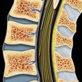
(Left) Sagittal graphic shows lumbar disc space infection with vertebral body osteomyelitis with endplate destruction and marrow edema. There are ventral and dorsal abscess collections.
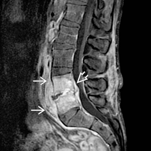
(Right) Sagittal T1WI C+ FS MR in this case of disc space infection shows enhancement of L5 and S1 bodies and intervertebral disc, with prevertebral and epidural phlegmon extension.
(Left) Axial T1WI C+ MR of a disc space infection shows inflammatory extension into the prevertebral space, psoas muscles, and dorsal spinal muscles. Phlegmon extends into the ventral epidural space with thecal sac compression .
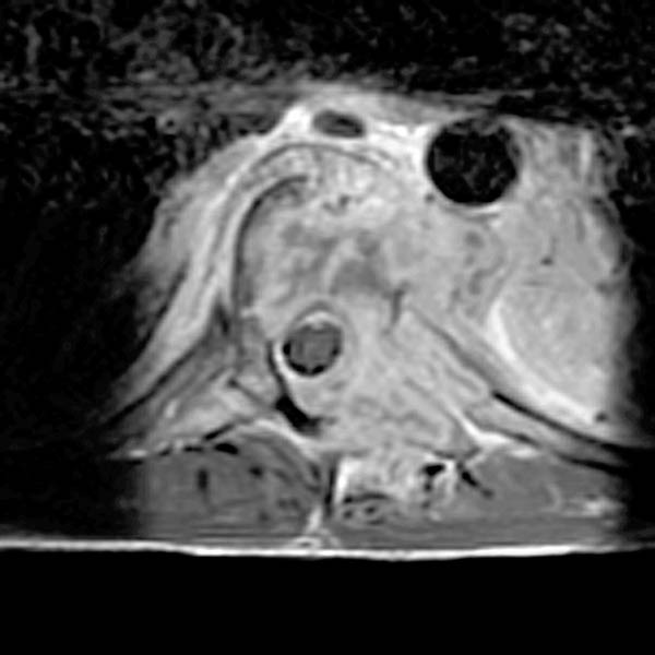
(Right) Axial T2WI FS MR shows inflammatory extension into the prevertebral space, psoas muscles , and dorsal spinal muscles .
(Left) Axial T1WI C+ MR shows disseminated coccidioidomycosis with diffuse bone and soft tissue involvement and adjacent paraspinal extension and extension into lung.
(Right) Axial T2WI MR in coccidioidomycosis shows huge paraspinal abscesses . There is effacement of the normal thecal sac within the spinal canal due to disc space infection and osteomyelitis.
(Left) Sagittal graphic shows dermal sinus extending from skin surface to conus, with conus abscess and extensive cord edema.
(Right) Sagittal T2WI MR in a patient with a cervical cord abscess and streptococcal endocarditis shows diffuse cord expansion, with a ring-shaped area of low T2 signal (abscess capsule) within the cord from C4 to C5-C6 .
Only gold members can continue reading. Log In or Register to continue
 and intervertebral disc, with prevertebral and epidural phlegmon
  extension.
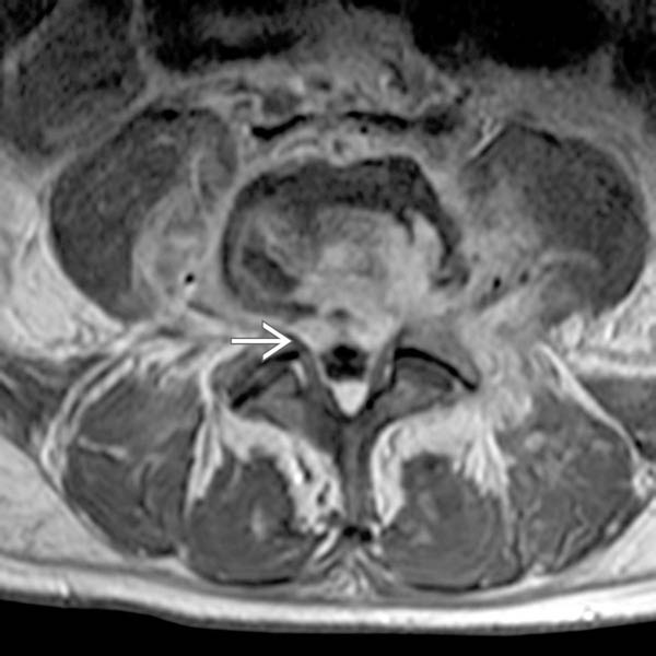
 .
.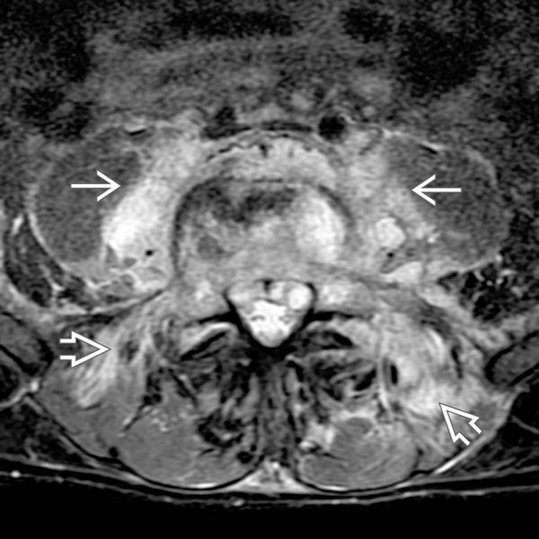
 , and dorsal spinal muscles
  .
.
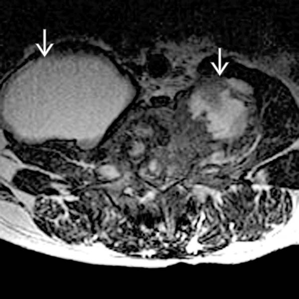
 . There is effacement of the normal thecal sac within the spinal canal due to disc space infection and osteomyelitis.
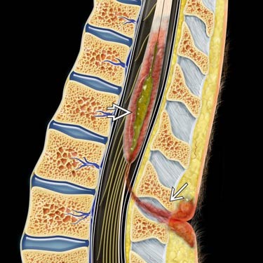
 extending from skin surface to conus, with conus abscess
  and extensive cord edema.
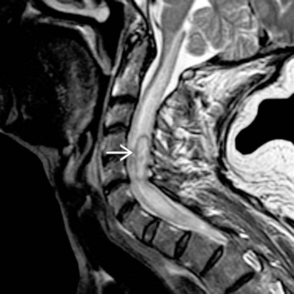
 .
.