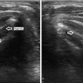
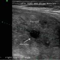
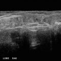
Fig. 17.1
Ultrasound images of squamous cell cyst show it is anterior to the strap muscles (a, right upper corner of image). In contrast, the normal thyroid is deep to the thin hypoechoic line of the sternothyroid muscle. Both the cyst and the thyroid have the same echogenicity. This relationship is not as evident in the transverse view (b) where the nodule appears to arise from the isthmus

Fig. 17.2
Excised benign squamous cell cyst and contents (a–c)
This patient’s imaging findings are interesting because they represent a benign superficial cyst mimicking a thyroid nodule, both in exam and ultrasound features. The case also illustrates the need for attentiveness to detail. While it may be instinctive to focus on the main abnormality, especially if it is a large mass, valuable information may come from slowing down the scanning pace to discern nuances such as spatial relationships to surrounding anatomical structures .
17.2 Case Vignette 2: Large Mass with Microcalcifications in the Central Neck
Two years following initial diagnosis and treatment for papillary thyroid cancer (PTC) at age 14, surveillance ultrasound was performed on a 16-year-old boy during routine follow-up visit. The referring physician noticed that the ultrasound report described a previously undetected mass in the central neck with microcalcifications and suspected recurrent PTC. Additional ultrasound imaging during consultation for possible biopsy and/or reoperation clarified that the mass had a different etiology: normal benign thymus. Neither biopsy nor surgery was performed. This case emphasizes that the thymus is a normal component of anatomy in the central neck, particularly in children and young adolescents. In this young population, the thymus typically has scattered hyperechoic foci that mimic microcalcifications (Video 17.4).
17.3 Case Vignette 3: Anaplastic Thyroid Carcinoma
This type of thyroid cancer remains one of the most aggressive, deadly malignancies of any kind. In this 62-year-old man with biopsy-proven anaplastic thyroid cancer, the large left-sided thyroid mass had developed rapidly over the course of 10 days. Normal anatomical structures were displaced laterally, as seen on an early computed tomography (CT) scan (Fig. 17.3). Placement of a tracheostomy tube was necessary for airway security and to allow chemoradiation and palliative care therapies to proceed. While the CT scan demonstrated how compromised and displaced the trachea was, it was impossible to palpate this by exam. Ultrasound played a key role to identify the location of the trachea and guide placement of a tracheostomy (Fig. 17.4).


