Fig. 12.1
An 18-month-old boy who underwent chest radiograph for evaluation of pneumonia in the setting of fever and elevated white blood cell count. Frontal chest radiograph demonstrates normal deviation (arrow) of the trachea to the right of midline at the thoracic inlet level
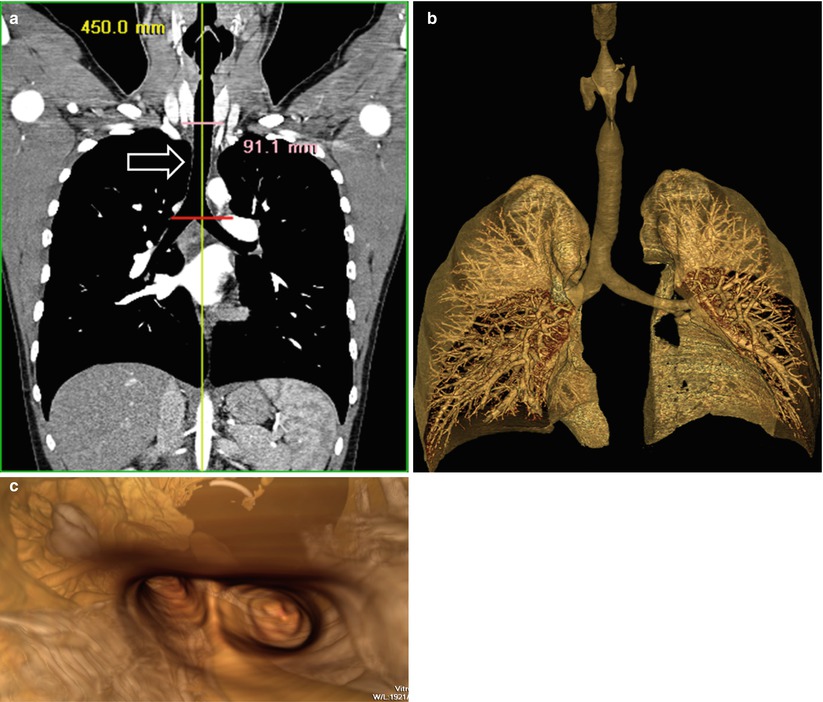
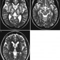
Fig. 12.2
Normal large airways of a 5-year-old girl. (a) Curved coronal reformatted CT image demonstrates a straightened view of the entire trachea (arrow). (b) Three-dimensional volume-rendered image of the patent large airways and lungs. (c) Three-dimensional volume-rendered image of the internal view (i.e., virtual bronchoscopy view) of the trachea facing the carina and the main stem bronchi shows patent central airways
12.5.2 Congenital Tracheal Stenosis
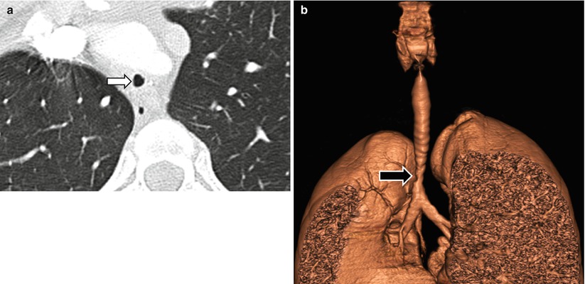
Fig. 12.3
Focal congenital tracheal stenosis in a 14-year-old girl who presented with stridor and wheezing. (a) Axial CT shows a narrowed trachea (arrow). (b) Three-dimensional volume-rendered image shows a high-grade short-segment tracheal stenosis (arrow)
12.5.3 Croup
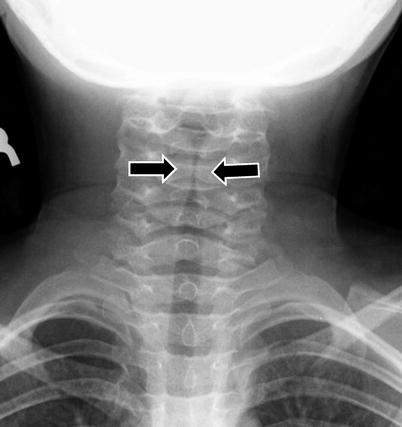
Fig. 12.4
Croup in a 2-year-old boy who presented with “barking cough.” Frontal radiograph shows symmetric subglottic narrowing (arrows), with loss of normal shouldering
12.5.4 Bacterial Tracheitis
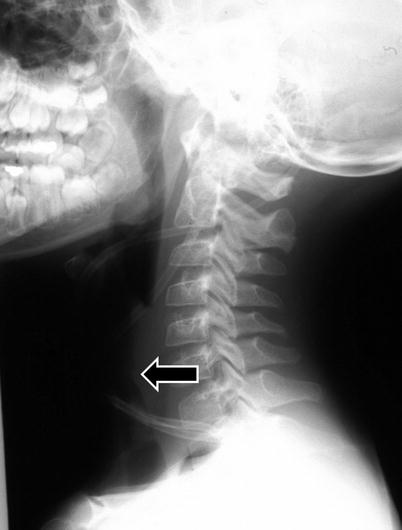
Fig. 12.5
Bacterial tracheitis in a 5-year-old boy with inspiratory stridor, cough and fever. Lateral neck radiograph shows tracheal irregularity and narrowing (arrow) (Reprinted with permission from American Journal of Roentgenology, page W167, 2013)
12.5.5 Epiglottitis
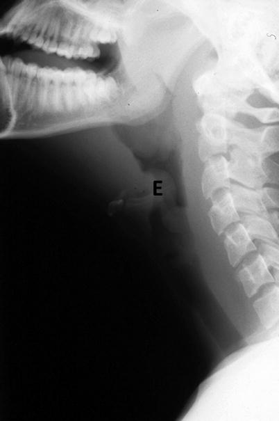
Fig. 12.6
Epiglottitis in a 6-year-old girl with fever, sore throat and drooling. Lateral radiograph shows enlarged epiglottis (E), which narrows upper airway (Reprinted with permission from American Journal of Roentgenology, page W168, 2013)
12.5.6 Tuberculosis
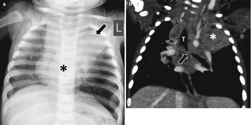
Fig. 12.7
Tuberculosis in a 6-month-old girl with a history of direct contact with active tuberculosis patient who presented with noisy breathing. (a) Frontal radiograph shows an opacity (asterisk) in the subcarinal region, likely representing an enlarged lymph node and left upper lobe confluent atelectasis (arrow). (b) Enhanced coronal CT image shows an obstruction (arrow) of the left main stem bronchus. Left upper lobe confluent atelectasis (asterisk) is again seen. T trachea
12.5.7 Subglottic Hemangioma
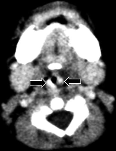
Fig. 12.8
Subglottic hemangioma in a 2-month-old boy who presented with worsening biphasic stridor. Enhanced axial CT image shows markedly enhancing subglottic mass (arrows), narrowing the airway at this level
12.5.8 Recurrent Respiratory Papillomatosis
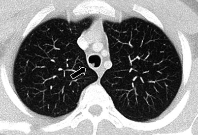
Fig. 12.9
Recurrent respiratory papillomatosis in a 17-year-old boy with known diagnosis since the age of 1.5 years. Axial CT image shows an intratracheal soft tissue lesion (arrow) (Case courtesy of Hedieh K. Eslamy, MD, Department of Radiology, Lucile Packard Children’s Hospital, Stanford, CA)
12.5.9 Carcinoid Tumor
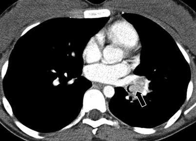
Fig. 12.10
Endobronchial carcinoid tumor in a 16-year-old girl who presented with recurrent left lower lobe pneumonia for the last 3 years. Enhanced axial CT image shows an intraluminal mass (arrow) involving the left lower lobe bronchus
12.5.10 Vascular Causes of Airway Abnormalities
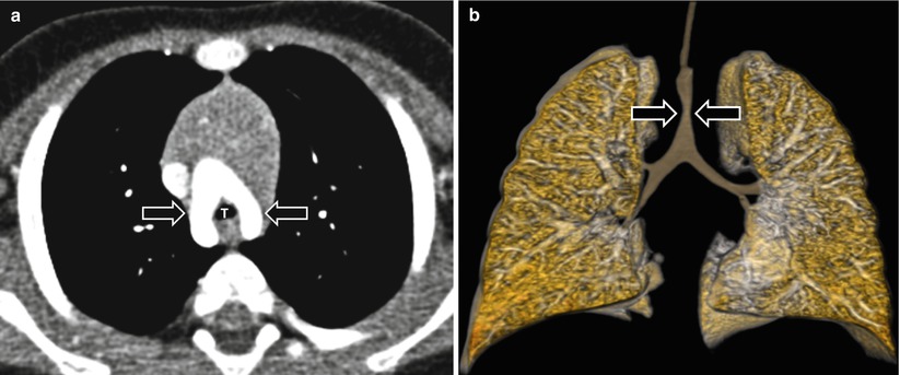
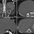
Fig. 12.11
Double aortic arch in an 8-month-old boy who presented with stridor, difficulty of feeding, and repeated apnea. (a) Enhanced axial CT image shows two (i.e., right and left) aortic arches (arrows) surrounding a trachea (T). (b) Three-dimensional volume-rendered image of the central airways and lung shows a tracheal narrowing (arrows) at the level of the double aortic arch
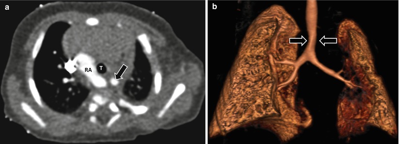
Fig. 12.12
Right aortic arch with an aberrant left subclavian artery in a 2 month-old girl who presented with respiratory distress and dysphagia. (a) Enhanced axial CT image shows a right aortic arch (RA) with an aberrant left subclavian artery (arrow). T trachea. (b) Three-dimensional volume-rendered image of the central airways and lung shows a mild tracheal narrowing (arrow)
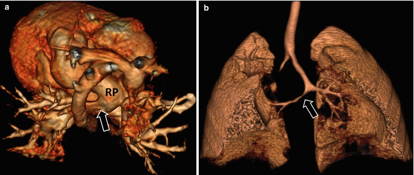
Fig. 12.13


Pulmonary artery sling in a newborn girl who presented with severe respiratory distress. (a) Three-dimensional volume-rendered image of the mediastinal vessels shows the left main pulmonary artery (i.e., pulmonary artery sling; arrow) directly arising from the right main pulmonary artery (RP). (b) Three-dimensional volume-rendered image of the central airways and lung demonstrates multiple congenital anomalies of the central airways, including a blind-ending right upper lobe bronchus, T-shaped carina, and right main stem bronchial stenosis (arrow)
Related posts:


Stay updated, free articles. Join our Telegram channel
Full access? Get Clinical Tree
