EU/CT/MRI: Caliceal dilatation and infundibular stenosis. Small renal pelvis.
US: Nonspecific. Caliceal dilatation. No renal pelvis dilatation.
Rare form of congenital hydrocalycosis. Dilated calyces drain through stenotic infundibula into a hypoplastic/stenosed renal pelvis. Different from infundibular stenosis, which has normal pelvis.
Fig. 3.39 EU in a young girl shows obstructed ureters on both sides displaced laterally by a pelvic “mass.” The extrinsic mass was found to be an enlarged uterus from hydrometrocolpos.
Table 3.13 Partial dilatation of the PC system
Caliceal diverticulum
EU/CT/MRI: Delayed opacification and drainage of diverticulum. Smooth margins and caliceal communication. May arise from any part of PC system.
US: cystic parenchymal lesion.
Congenital or acquired. Typically asymptomatic, but may be complicated by infection or stones.
Upper caliceal dilatation
CECT/MRI: demonstrates caliceal dilatation and cause.
US: Echogenic mass in collecting system. No Doppler signal.
EU: CT irregular; castlike filling defect in the collecting system.
Candida is most common infection. Usually an opportunistic infection in the immunocompromised patient.
Fibroepithelial polyp
EU: irregular filling defect attached by a stalk at one end to the ureter.
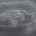
Fig. 3.40 Right renal staghorn calculus. The shape of the calculus conforms to the collecting system.Fig. 3.41 Pyonephrosis. US image of kidney shows dilated collecting system with layering echogenic debris.Fig. 3.42 Pyonephrosis. US image of the kidney in another child with pyonephrosis shows dilated calyces with particulate internal echoes and debris.
US: thin-walled cystic structure in bladder, extending from ureter.
CT/EU: cobra head appearance of orthotopic ureterocele.
VCUG: filling defect in bladder on early filling films.
Ectopic ureteroceles: Associated with upper pole of duplex kidney. More common in females (6 times more common).
Orthotopic ureteroceles: Rarer. Often incidental findings in older children/adults. Normal position at trigone.
Ectopic (subvesical) ureter
Dilated ureter and dysplastic upper moiety of duplex system may be shown on all modalities.
EU/CT/MRI: can show ectopic insertion.
VCUG: reflux in 30%.
Presentation: dribbling in girls, epididymo-orchitis in boys. Sixty-eight to 80% are associated with duplication, where the ectopic ureter inserts caudal and me-dial to the normal ureteric insertion (Weigert-Meyer law). May drain into lower bladder, urethra, vestibule, or vagina in girls; lower bladder, posterior urethra, seminal vesicles, or vas deferens in boys.
Functional dilatation of the ureter
US/CT/MRI: dilated ureter with no intraluminal obstruction.
May be seen with postobstructive diuresis, diabetes insipidus.
Ureteral stump
US: stump may appear as diverticulum or blind ending ureter.
CT/MRI: may show reflux into stump on delayed imaging.
Results from heminephrectomy of duplex system or nephrectomy from any cause.
Retrocaval ureter
EU/CT/MRI: Medial deviation of ureter at L3 level, passing behind ureter. Classic sigmoid or “fish-hook” appearance.
Almost always right-sided. Results from persistence of the posterior cardinal venous system.
Ureteral dysplasia
US/CT/MRI: Very dilated tortuous ureters with absent peristalsis. May show dysplastic kidneys.
VCUG: gross reflux.
Histology shows poor muscularization of ureteral wall, leading to absent/poor peristalsis. Association of renal dysplasia is less than expected.
Fig. 3.43 Ureterocele. VCUG image shows a filling defect in the posterior urethra caused by prolapsed ureterocele of an ectopically inserted ureter.
Table 3.16 The enlarged bladder
Diagnosis
Findings
Comments
Functional disturbance of bladder emptying
US: Large capacity but normal appearing bladder. Large residual.
VCUG: no reflux.
Causes: status postcatheterization, intervention, analgesia, infection, trauma.
Infrequent voider, lazy bladder syndrome
Large but otherwise normal bladder with large residual.
US: may have prominent upper tracts.
VCUG: urodynamics show hypotonic bladder, prolonged decreased voiding.
No/infrequent urge to void every 8–12 h. Incontinence. Frequent urinary tract infections. Associated with constipation.
VCUG: Bladder wall trabeculation. Stricture and dilated proximal segment of urethra noted. Ascending urethrography shows true distal extent of stricture.
Causes: usually iatrogenic (catheterization and cystoscopy), but also trauma, foreign bodies, extrinsic compression from pelvic floor tumors.
Neuropathic bladder
Abdominal X-ray: may show spinal dysraphism, sacral agenesis, spinal trauma.
US/VCUG: Thick-walled, trabeculated, or large atonic bladder depending on level of lesion. Pseudodiverticula. Elongated funnel-shaped posterior urethra on voiding.
Suprasacral lesions give an elongated pine cone appearance to the bladder. Suprapontine lesions give a rounded serrated appearance. Peripheral (below S2–S4) lesions demonstrate a large atonic bladder.
Vesicourachal diverticulum is an outpouching at the apex of the bladder resulting from incomplete closure of the proximal urachus.
The majority of patients are asymptomatic because the diverticula drain well with bladder emptying.
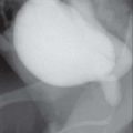
Fig. 3.44 Bladder diverticulum. Cystogram during VCUG demonstrates a smooth-walled posterior bladder diverticulum.Fig. 3.45 Bilateral reflux on VCUG image. A diverticulum is present in relation to the right VUJ, which is typical of a Hutch diverticulum.Fig. 3.46 A posterior urethral valve resulting in dilatation of the posterior urethra.Fig. 3.47 Urethral strictures. (a, b) Multiple urethral strictures are seen on cystourethrogram causing focal narrowing and dilatation of the membranous and posterior urethra.Fig. 3.48 Urethral stenosis causing urethral dilatation in a girl. Note the vaginal reflux, which is common during cystourethrography in the female child.Fig. 3.49 Urachal diverticulum.
VCUG: Diff cult to catheterize. Pinpoint meatus on voiding. Trabeculated bladder with residual.
Pinpoint appearance of meatus on visual inspection. May be congenital or secondary to surgery (circumcision/hypospadias) or trauma.
“Spinning top” urethra
VCUG: triangular appearance of the urethra in girls.
Associated with bladder instability.
Wide bladder-neck deformity
Table 3.18 Filling defects in the urethra
Diagnosis
Findings
Comments
Posttraumatic
VCUG/retrograde urethrography: Acutely, will show contrast extravasation at site of injury; evaluate bladder. Later, will get irregular polypoid changes in the lumen.
MRI: can assess urethra and distinguish type IV from type IVa injuries.
More common in males. Goldman classification from type I to V.
Polyp on stalk in proximal urethra, extending into the bladder. Obstruction is rare.
Similar to ureteral polyp. Benign fibroepithelial hyperplasia.
Fig. 3.50 Filling defect. Cystogram demonstrates a well-defined filling defect at the base of bladder on the right side from a prolapsed ureterocele.Fig. 3.51a, b Urethral polyp. Cystourethrogram shows a lucent filling defect within the urethra caused by a urethral polyp.
Table 3.19 Calcifications in the renal fossa
Diagnosis
Findings
Comments
Nephrolithiasis
US: Echogenic foci with posterior shadowing. Doppler may show twinkle sign.
(see unilateral large kidney without PC dilatation, Table 3.4)
Oxalosis
US/EU/CT: widespread faint medullary calcification, with early stone formation.
Can be inherited or secondary to extensive small bowel resection.
Acquire osteosclerosis.
After renal trauma
Calcification of a perirenal hematoma.
Fig. 3.52a, b Renal pyramids. (a) US image shows hyperechoic pyramids in a child with nephrocalcinosis. (b) Abdominal radiograph shows calcifications in both renal areas, conforming to the renal pyramids.
Table 3.20 The small capacity bladder
Diagnosis
Findings
Comments
Detrusor hyperreflexia instability
US: Normal or small bladder with wall thickening. Normal upper renal tracts.
VCUG: Small, hypertonic bladder, with intermittent widening and contraction of bladder neck. VUR may be seen.
Increased day time frequency; normal urine examination. benign and self limiting condition.
Cystitis
Plain X-ray: bladder calculi; intramural or intraluminal air in emphysematous cystitis.
US: small capacity bladder with irregular thick walls (> 3.5 mm), internal echoes, postvoid residual urine.
CT: may show bladder calculi, diverticula, fistula formation, or pelvic abscess with complicated cystitis.
MRI: small capacity bladder with wall thickening, increases T2 signal in wall, and enhancement with gadolinium.
Acute hemorrhagic cystitis, cyclophosphamide-induced and tumoral cystitis are the most important forms of pediatric cystitis. Correlation with urine examination is essential.
Neuropathic bladder
Plain X-ray: may show associated spinal anomalies, calculi.
US/VCUG: thick-walled trabeculated bladder with or without upper tract dilatation (from VUR); abnormal bladder shape—Christmas tree type (elongated) or atonic type (with diverticula or pseudodiverticula).
Urodynamic studies may confirm elevated intravesical pressures, detrusor sphincter dyssynergia. MRI is usually done to evaluate associated neurologic abnormalities.
Status postbladder surgery
US: small capacity, irregular outline, with significant postvoid, postoperative collections.
VCUG: extravasation or fistula formation.
CT/MRI: pelvic collections or fistulae.
Bladder rupture
US: free fluid in pelvis.
CT: Pelvic fractures in case of trauma. Delayed CECT images or CT cystography shows extravasated contrast (in paracolic gutters and interloops in case of intraperitoneal rupture or perivesical space and perineum and upper thighs with extraperitoneal rupture). Combined intra- and extraperitoneal rupture may occur.
Retrograde ureteral study shows associated urethral rupture.
Intraperitoneal rupture occurs at the dome, which is weakest part.
Extraperitoneal rupture is close to bladder base antero-laterally.
Bladder exstrophy, status post surgery
(see status postbladder surgery)
US: Small bladder capacity, wall thickening, upper tract dilatation, calculi (due to urinary stasis). Postoperative collections.
Scrotal US for epididymitis as a late complication.
NM: DMSA for renal cortical scarring.
Surgery includes closure of bladder exstrophy, bladder neck reconstruction, and epispadias repair in a staged manner with or without bladder augmentation. Appendicovesicostomy allows for percutaneous bladder catheterization. VUR is seen almost always postoperatively.
Bilateral ectopic ureters
US: small capacity bladder with dysplastic kidneys.
VCUG: associated reflux.
EU/CT or MRI urography abnormal course and ectopic insertion of ureters
US: Lobulated, polypoid mass usually from trigone > bladder neck > dome. Local spread to lymph nodes. MRI for extent.
Most common neoplasm of urinary tract. Occurs most often in bladder but can occur in prostate or vagina. Embryonal type most common. Age < 5 y. M > F. Diagnosis confirmed with biopsy.
Hemangioma
US: Discrete mass usually arising from bladder dome. Doppler imaging for assessment of vascularity. Calcifications may be present.
CT: May show vascular mass enhances with contrast.
MRI: better for characterization and evaluation of extent.
Most common benign bladder tumor. Coexisting cutaneous hemangioma over lower abdomen, pelvis, or thigh may be found.
Venolymphatic malformations may also present as filling defects in bladder although they p resent c ommonly with hematuria. Mulitiple visceral hemangiomas, rectal varices, and genitourinary venolymphatic malformations may be seen in Klippel Trenauney syndrome.
Fig. 3.53 Bladder hematoma. Cystogram shows a distended bladder opacified by contrast, with a large filling defect.Fig. 3.54a, b Intraperitoneal rupture of the bladder. (a) US image of the urinary bladder clot seen as mobile echogenic material within the lumen. (b) Cystogram in a child who sustained pelvic fractures shows contrast extravasation, confirming intraperitoneal rupture of the bladder.Fig. 3.55 Vesical calculi. Abdominal radiograph demonstrates two well- defined large radiopaque vesical calculi.Fig. 3.56a, b Rhabdomyosarcoma. Lobulated echogenic mass at the base of bladder and the prostate.
VCUG: oval contrast–filled outpouching along ventral aspect of anterior urethra.
High-resolution ultrasonography: shows the diverticulum if filled and the anterior lip.
May be congenital or acquired (secondary related to trauma, catheter, etc.).
Prune belly syndrome
VCUG: large bladder ± VUR, dilated high placed posterior urethra.
US: large bladder, dilated posterior urethra, hydro-nephrosis/dysplastic kidneys.
Predominantly seen in males. Triad of deficient anterior abdominal wall musculature, undescended testes, and urinary tract anomalies, particularly dysplastic kidneys and dilated posterior urethra.
VCUG: Demonstrates partial or complete urethral duplication.
Retrograde urethrogram (RGU) can be used for diagnosis.
Partial duplication can be proximal or distal. Can be epispadiac or hypospadiac depending on the position of accessory external openings.
Megaurethra
VCUG: Marked dilatation of penile urethra with proximal and distal tapering into normal caliber urethra. There is no obstruction.
US: may show deficiency/absence of corpus spongiosum and corpora cavernosa.
May be associated with prune belly syndrome or can present as an isolated anomaly. Can be of scaphoid or fusiform shape; scaphoid is more commonly seen as a ventral bulge of penile urethra during voiding.
Only part of anterior urethra may be affected.
Status postepispadias repair
VCUG/RGU: Postoperative strictures and prestenotic dilatation. Urethro- or vesicocutaneous fistulas if present.
Lacuna magna
VCUG/RGU: displays the lesion as dorsal and parallel to distal anterior urethra.
Dorsal diverticulum in the roof of fossa navicularis. Approximately 5 mm in size. Rarely symptomatic. Pain postvoid spotting or hemorrhage may be present.
Fig. 3.57 Urethral diverticulum. Contrast-opacified outpouching from the anterior urethra resulting from urethral diverticulum.Fig. 3.58a, b Urethral duplication. Cystourethrogram in a child with urethral duplication. Note a dilated posterior urethra and the accessory anterior urethral channel of a smaller caliber.
Only gold members can continue reading. Log In or Register to continue